
Стоковая фотография 1459919189 Pedis X Ray Anatomy Radiology Radiographic Shutterstock
The standard x-ray examination of the chest consists of a frontal (PA) and lateral view. The frontal view is called a PA view because the patient stands with the anterior chest on the cassette and the back to the x-ray beam. The x-rays first hit the posterior and then the anterior chest before hitting the cassette; thus the name PA. The cassette is 6 feet from the x-ray tube. The lateral film.
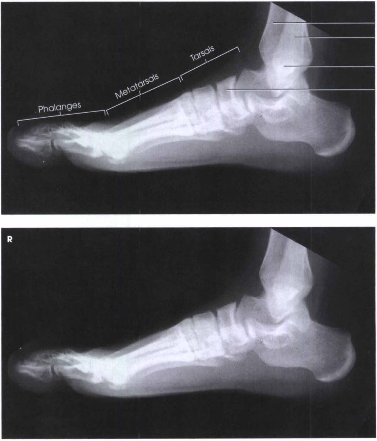
NORMAL FOOT 7
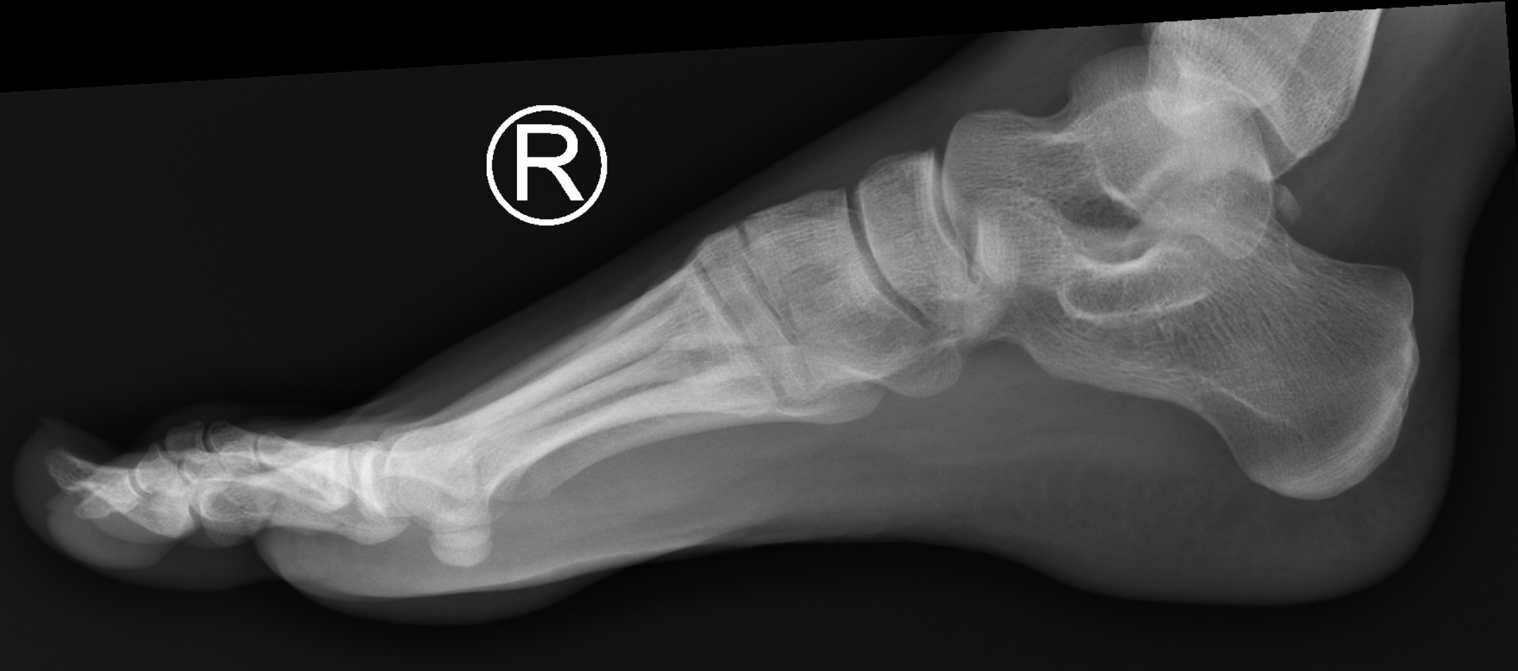
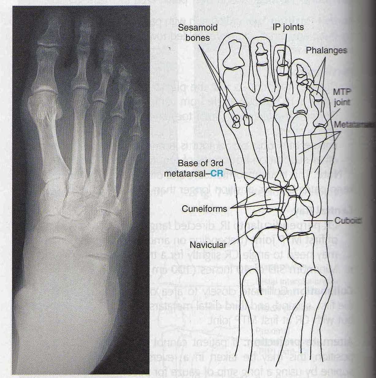
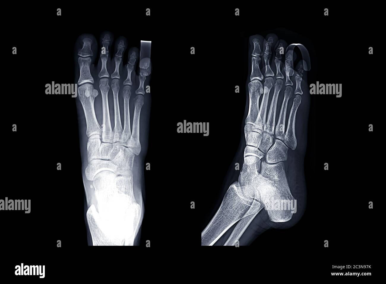
Imaging protocols have to consider the patient's comfort, level of anxiety, and smaller size. The first imaging study is usually made with plain radiographs. The routine radiographic examination of the foot includes the anteroposterior (AP), lateral, and oblique projections. Magnetic Resonance Imaging (RMI) provides excellent anatomic detail.
Gudang Medis teknik radiografi pedis
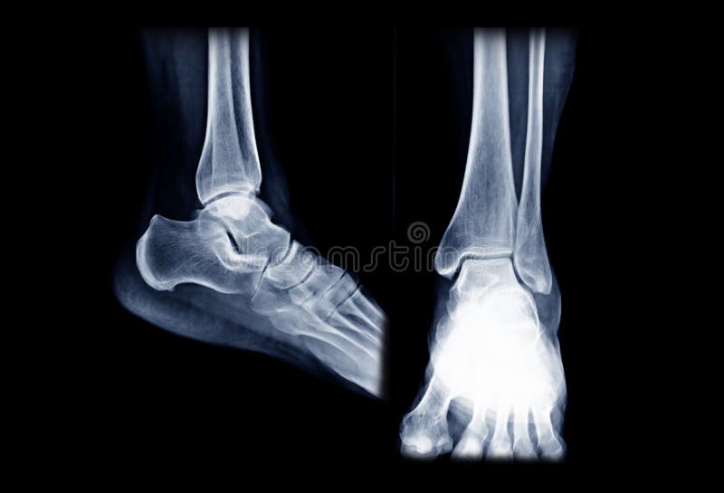
the lateral and medial malleoli of the distal fibula and tibia are in profile. the tibiotalar joint space should be open, yet the full mortise joint should not be visualized on the AP. Practical points. This view can be thought of as the literal anteroposterior of the ankle. Most patients will naturally place their foot in this position.
Gudang Medis teknik radiografi pedis
Selamat datang di RS Mutiara Bunda. 0726 7721215/ 08136916722 24 JAM (Senin-Minggu) [email protected].

Plain radiograph (AP and lateral oblique) of the left foot (injured)... Download Scientific
The answer is… below. On the PA view, the cardiac borders are smaller and more defined. Given the way the x-ray beam works, the heart appears smaller and with sharper borders on the PA view. The reason is that the patient's chest (anterior) is against the x-ray film with the beam entering from posterior (P) to anterior (A) - hence the.

AP and lateral left foot Xray, showing sclerotic of bone deformity... Download Scientific Diagram
(dorsoplantar) and lateral views of a skeletally immature right foot with normal forefoot and hindfoot alignment. Figures 1C and 1D show a younger child with normal anteroposterior and lateral foot findings for comparison. The tarsal bones are in - completely ossified, but the relationships of the talus and cal -
Xray Image of Right Ankle Joint AP and Lateral View. Stock Photo Image of examination, human
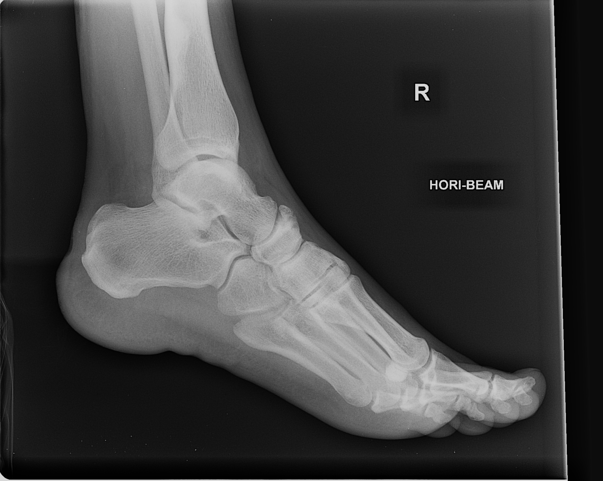
For the lateral view, the unaffected limb is placed behind the patient with the affected side down. Support can be placed along the distal aspect of the affected foot to obtain a true lateral image. Knee: For the AP view of the knee, the leg is extended and centered to the central ray. The leg is rotated slightly inward in order to place the.
Teknik Radiografi Ossa Pedis RADIOLOGI
the entire lumbar spine should be visible, with demonstration of T11/T12 superiorly and the sacrum inferiorly. no patient rotation as evident by central spinous processes and the symmetrical appearance of the sacroiliac joints and iliac wings. adequate image penetration and image contrast is evident by clear visualization of lumbar vertebral.

Álbumes 104+ Foto Radiografia De Pie Ap Y Lateral Lleno
Fleksibilitas dari lateral complex membuat talus dan fibula bergerak dan berputar selama pergerakan normal dari ankle. Pergerakan fibula ini pada syndesmosis merupakan bagian penting dari fungsi normal ankle. Teknik Radiografi. Proyeksi yang sering digunakan pada pemeriksaan ankle joint adalah AP dan Lateral.
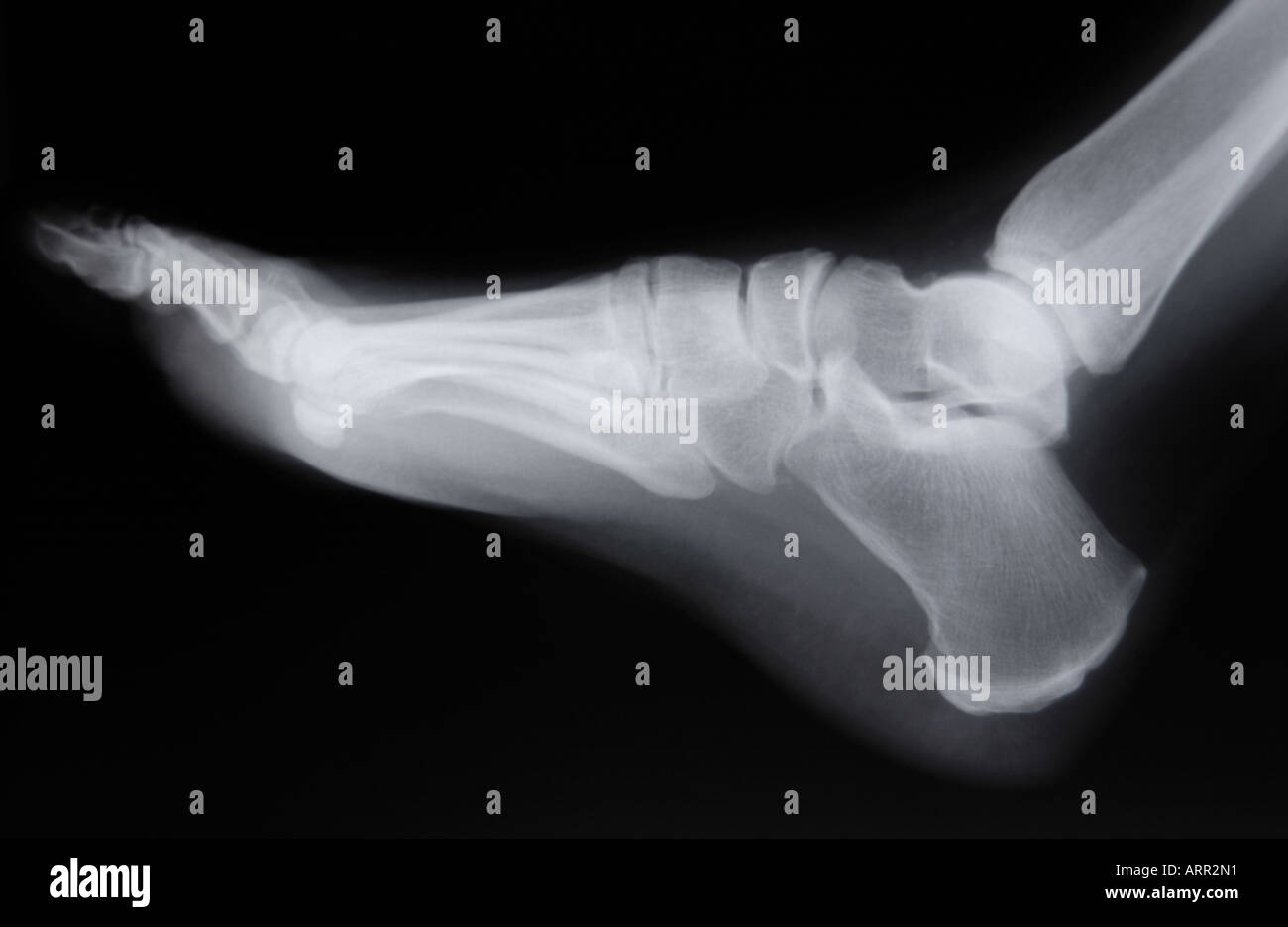
normal lateral xray of adult foot Stock Photo Alamy
Detail Layanan : Pemeriksaan Menggunakan X-Ray untuk melihat Gangguan pada Fungsi dan Struktur Daerah Dada (Ap/Lat 1x) Pasien akan diminta untuk membuka pakaian ketika melakukan pemeriksaan ini. Jika anda wanita dan sedang Hamil maka mohon menginformasikan kepada Petugas atau Konsultasi dengan Dokter Kandungan anda sebelum melakukan tes ini.
Normal calcaneal radiographs Image
Practical points. The fibula head is a great indication of rotation, if the fibula head is entirely superimposed, the image is not AP; to correct this you must internally rotate until the knee is in even contact with the image detector. Very slim patients may require a slight caudal angle to better visualize the joint space in an AP fashion.
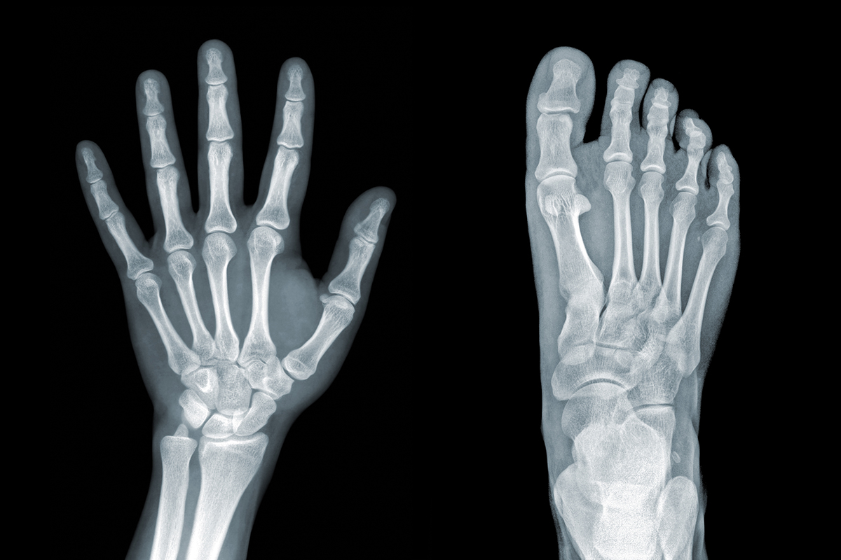
Orthopaedics foot/ankle and wrist/hand
AP radiograph shows that the tip of the fibula is abutting the lateral margin of the. Aykanat F, Vincenten C, Cankus MC, Kose O, Sindel M. Lateral foot pain due to os vesalianum pedis in a young football player; a case report and review of the current literature. Skeletal Radiol. 2019;48(11):1821-8. Article PubMed Google.
Comparaison de l'image du pied droit AP et de la vue oblique avec la sprint de doigt pour
Proyeksi : AP . 1.Kaset : ukuran 24 x 30 cm. 2. kV : 60 ± 5 mAs : 2. 3. FFD : 100 cm. 4. Posisi Pasien : Pasien tidur di atas meja pemeriksaan, taruh bantal di kepala, lulut di tekuk flexi dan telapak kaki berada di atas kaset. 5. Posisi Obyek : Tempatkan telapak kaki lurus dan rata di atas permukaan kaset. Kaki sejajar dengan kaset dan CR. 6.
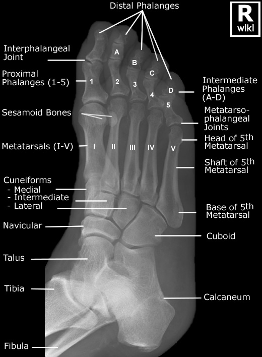
Foot Radiographic Anatomy wikiRadiography
2012 ICD-9-CM Procedure Code 87.1. Other X-Ray Of Face, Head, And Neck. A child code below 87.1 with greater detail should be used. 2012 ICD-9-CM Procedure Code 87.11. Full-Mouth X-Ray Of Teeth. 87.11 is a specific code and is valid to identify a procedure. 2012 ICD-9-CM Procedure Code 87.12. Other Dental X-Ray.

Normal radiographic anatomy of the foot Radiology Case Sănătate
Persiapan Pasien. Rontgen ankle dan kaki tidak memerlukan persiapan khusus. Secara umum, persiapan meliputi anamnesis dan pemeriksaan fisik untuk menentukan kecurigaan posisi lesi dan memilih seri rontgen yang sesuai. Setelah itu, berikan penjelasan lengkap mengenai indikasi prosedur dan langkah-langkahnya.

Pedis X Ray Anatomy Radiology Radiographic库存照片1459919177 Shutterstock
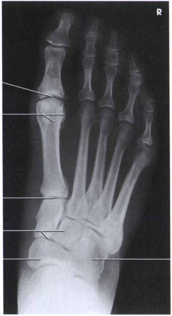
First cuneiform (medial) Medial sesamoid. Lateral sesamoid. Shaft of second metatarsal. Neck of third metatarsal. Head of fourth metatarsal. Metatarsophalangeal joint. Proximal phalanx. Middle phalanx.