ECG Lead positioning • LITFL • ECG Library Basics
Ada enam sistem lead ekstremitas, yaitu tiga lead "bipolar" yang berasal dari segitiga Einthoven dan tiga sistem lead "unipolar" yang dikembangkan pada tahun 1940-an.. Ada situasi tertentu di mana menempatkan semua 10 elektroda untuk EKG 12-lead bisa jadi sulit. Untuk menanggapi kebutuhan ini, sistem empat kabel yang lebih sederhana.

ECG Lead positioning • LITFL • ECG Library Basics
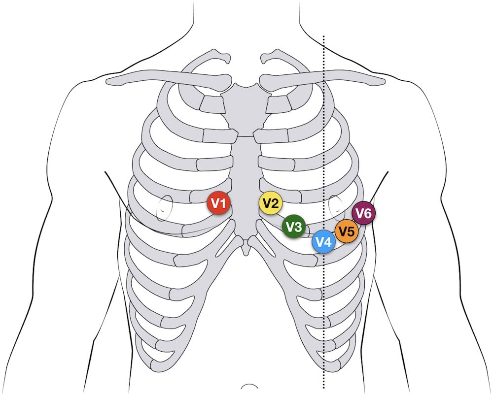
ECG chest (precordial) lead placement - left, anterior oblique view . Finding the Correct Placement of Leads V1 - V6 Placement of Lead V1. Locate the sternal notch (Angle of Louis) by feeling the top portion of the breast bone, and moving your fingers downward until you feel a bump. Move your fingers to the right, off of the bump, and you.

Ilustración De Posición Del Electrodo De Ecg Con Pistas De Extremidad De Ecg. útil Para Educar a
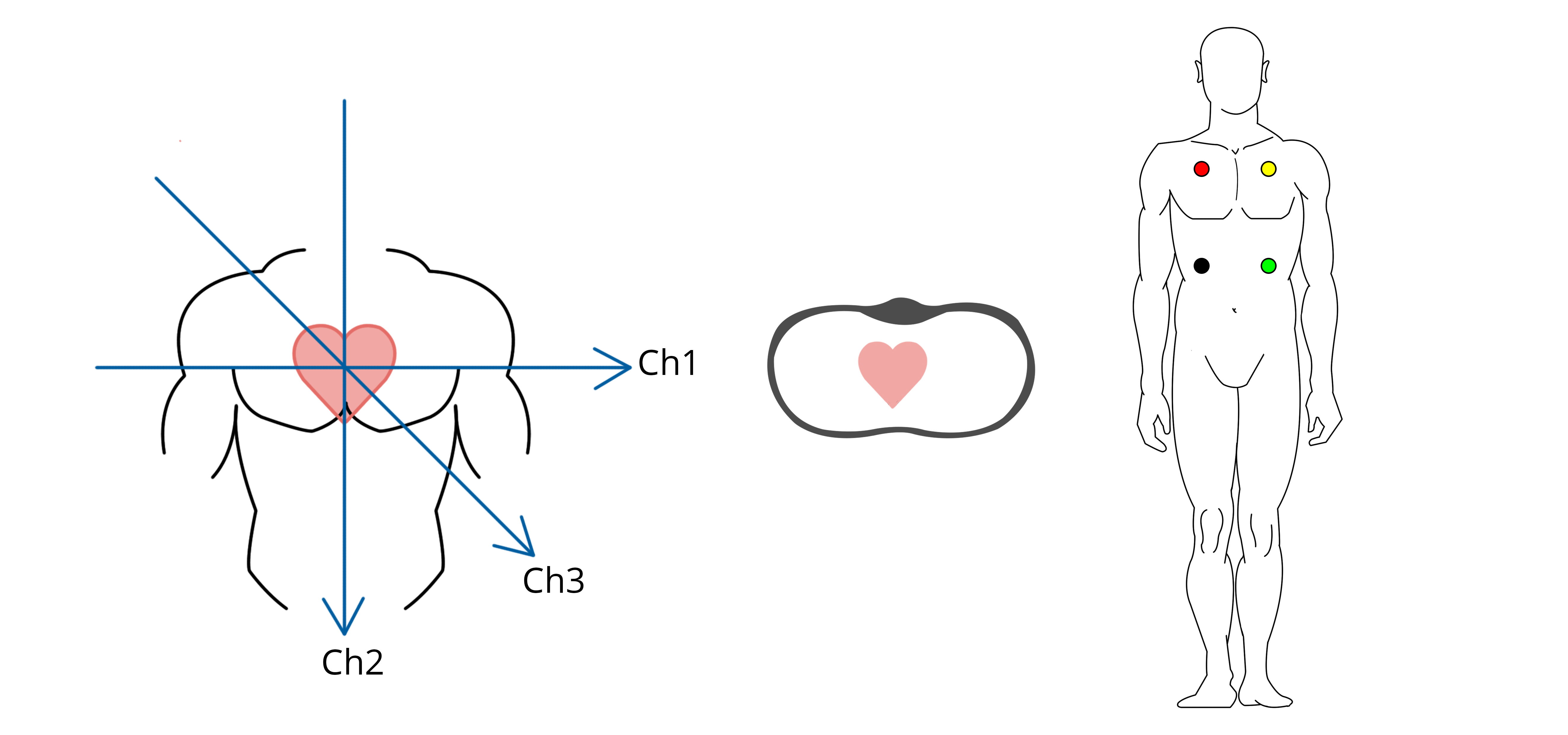
Electrodes. ECG electrodes are pads that attach to the skin, conduct electricity and connect to the ECG machine to generate the 12 leads that detail the patient's heart activity. While there are 12 leads, there are only ten electrodes. With a standard 12-lead ECG, six chest electrodes and four limb electrodes adhere to the skin.
Lead systems how an ECG works CardioSecur
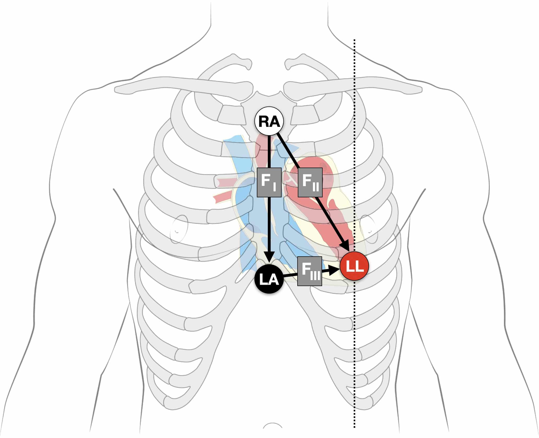
Elektrode ekstremitas atas dipasang pada pergelangan tangan kanan dan kiri searah dengan telapak tangan.. Dengan memindahkan lead selector kemudian dibuat pencatatan EKG secara berturut-turut yaitu sandapan (lead) I, II, III, aVR, aVL, aVF, VI, V2, V3, V4, V5, V6. Setelah pencatatan, tutup kembali dengan kalibrasi seperti semula sebanyak 2-3.

12 Lead Electrocardiogram (ECG) in Australia Dr Arthur Nasis
An ECG electrode is a conductive pad attached to the skin to record electrical activity. The data gathered from these electrodes allows the 12 leads of the ECG to be calculated (e.g. lead I is calculated using data from the electrodes on both the right and left arm ). The electrodes used to generate a 12-lead ECG are described below.
ECG Lead positioning • LITFL • ECG Library Basics
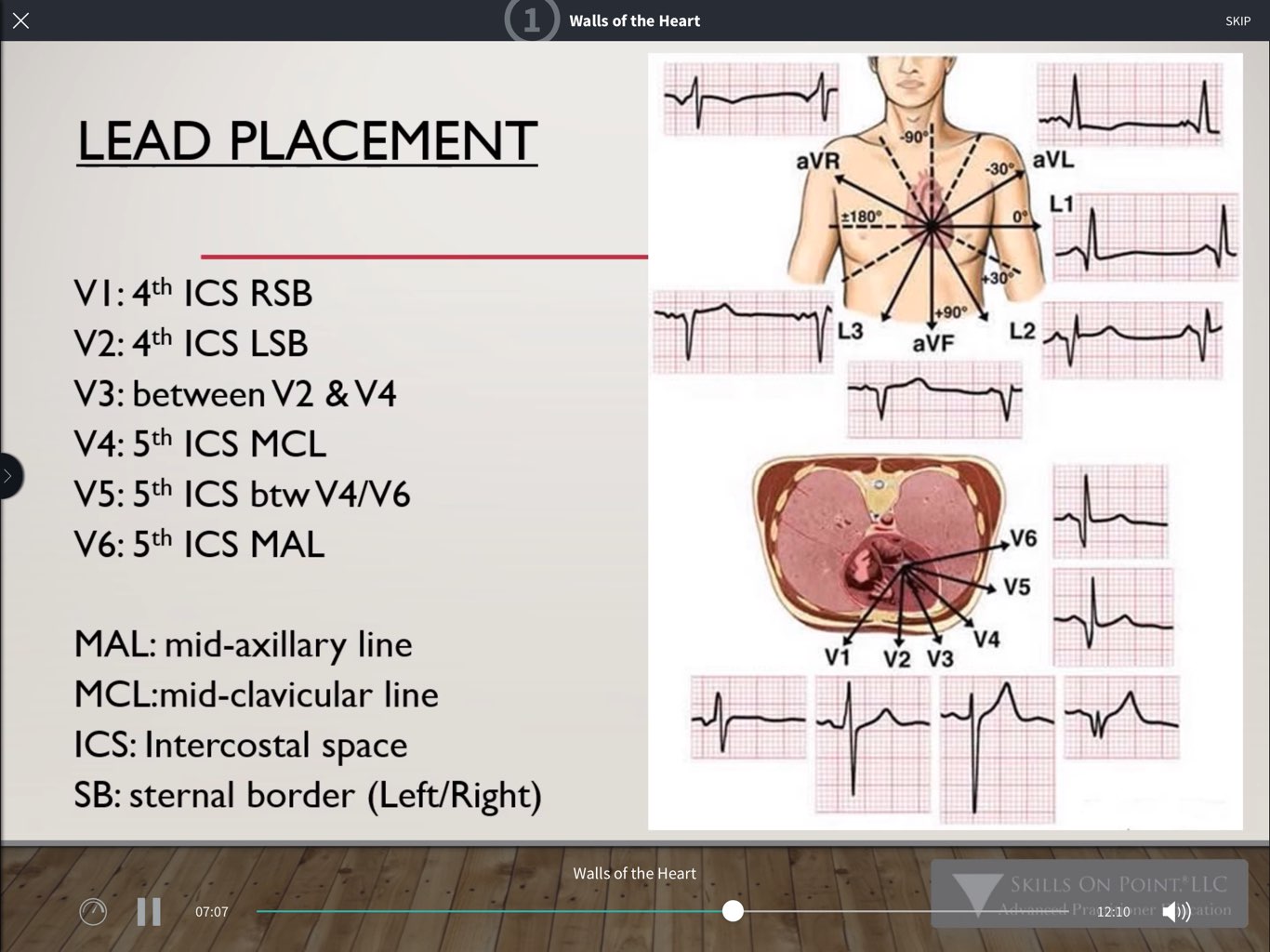
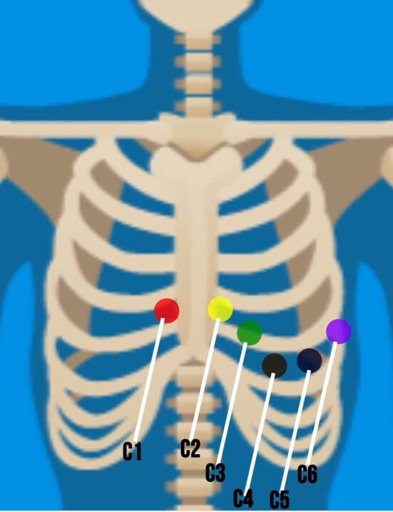
V1: this chest lead registers potentials from the atria, part of the septum and the right ventricle anterior wall. The QRS complex is formed by a small R wave (septum depolarization) followed by a deep S wave (ventricles activation), see QRS morphology.; V2: the electrode for this precordial lead rests over the right ventricle wall. Therefore, the R wave is slightly bigger than in V1, followed.
Lead systems how an ECG works CardioSecur
Introduction. Patients frequently consult their general practitioner (GP) with symptoms that may indicate an underlying cardiac arrhythmia (1,2).When a cardiac arrhythmia is suspected, a 12-lead electrocardiogram (12L-ECG) is indicated ().Due to logistical challenges, only a third of ECGs is performed while a patient is experiencing symptoms ()..
Elektrokardiografi (EKG) doktermedis
ECG monitoring is common place in the hospital and even pre-hospital setting. The need for different types of lead systems in different settings has been emphasised. Simple three electrode bipolar recording is ubiquitous for monitoring. This can be used to record modified bipolar chest leads as well. Using five leads gives the option of getting.

5Lead ECG Interpretation (Electrocardiogram) Tips for Nurses FRESHRN
ECG (EKG) Interpretation. As with all investigations the most important things are your findings on history, examination and basic observations. Having a good system will avoid making errors. To start with we will cover the basics of the ECG, how it is recorded and the basic physiology. The 12-lead ECG misleadingly only has 10 electrodes.

[DIAGRAM] Standard 12 Lead Ecg Placement Diagram
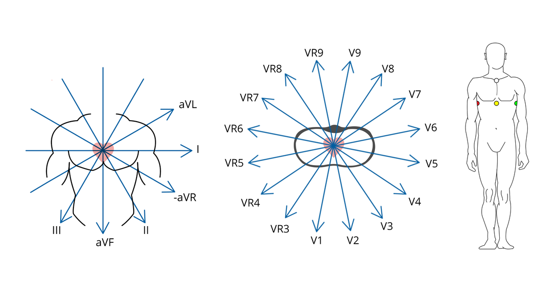
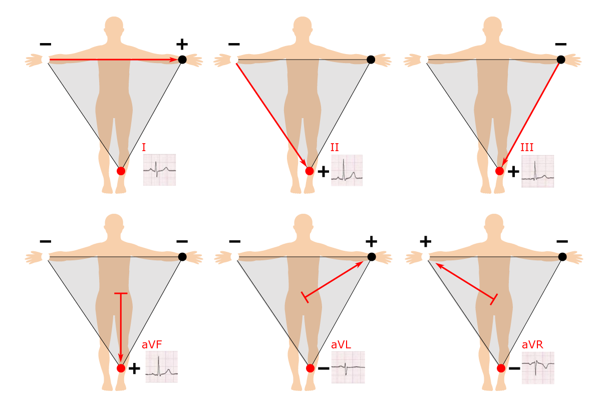
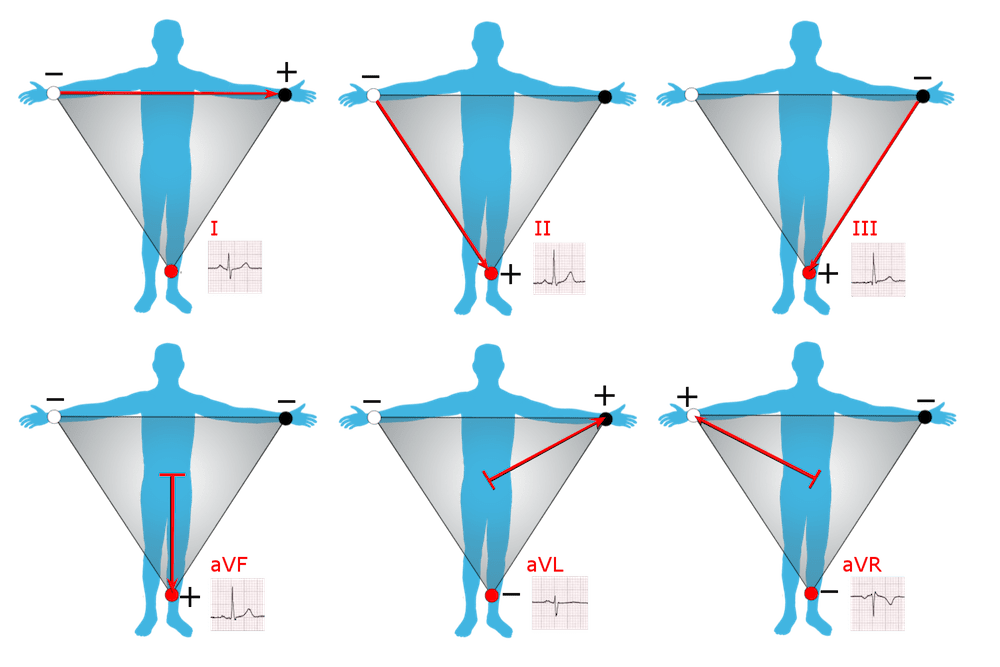
EKG 12 Lead. Lead I, aVL, V5, V6 menunjukkan bagian lateral jantung. Lead II, III, aVF menunjukkan bagian inferior jantung. Lead V1 s/d V4 menunjukkan bagian anterior jantung. Lead aVR hanya sebagai petunjuk apakah pemasangan EKG sudah benar. Aksis jantung. Sumbu listrik jantung atau aksis jantung dapat diketahui dari bidang frontal dan horisontal.
12Lead ECG Placement Guide with Illustrations
Pericarditis is classically associated with ECG changes that evolve through four stages. Stage 1 - widespread STE and PR depression with reciprocal changes in aVR (occurs during the first two weeks) Stage 2 - normalisation of ST changes; generalised T wave flattening (1 to 3 weeks) Stage 3 - flattened T waves become inverted (3 to several.

ECG Lead positioning • LITFL • ECG Library Basics
Here are the practical EKG parameters to remember when doing 12 lead EKG interpretation. EKG Measurements and Calibration. The smallest box on the paper = 1 mm on all sides. A big box containing 5 small boxes = 5 mm on all sides. Measuring Time. Time is measured horizontally. 1 mm = 0.04 sec = 40 ms. 5 mm = 0.2 sec = 200 ms. 25 mm = 1 sec.
Basic 12 Lead EKG Interpretation Complete Anatomy
The 2020 ESC AF management guideline states that a 30-second single-lead electrocardiogram (sl-ECG) can be used to diagnose atrial fibrillation, but advises caution as most validation studies used small, carefully selected cohorts and are prone to bias. Patients with frequent ectopy, other atrial arrhythmias, and cardiac implantable electronic.
ECG Limb Lead Reversal • LITFL • ECG Library Diagnosis
Teknik. Teknik pemeriksaan elektrokardiografi atau EKG harus diperhatikan untuk memastikan hasil pemeriksaan yang akurat. Prosedural pemasangan elektroda merupakan salah satu bagian teknik EKG yang penting agar artefak yang muncul hanya minimal, atau tidak sama sekali. Persiapan pasien untuk EKG mencakup informed consent pasien, cek identitas.

12 Lead ECG Electrode Placement
A posterior STEMI is also generally not visible on the 12 lead EKG, as it is measured by leads V7-9 which are not applied during a standard 12 lead EKG. Much like leads V1-6 wrap around the left side of the chest, leads V7-9 continue to wrap around the posterior section of the chest wall. We will not discuss leads or placement beyond the.
Cara Pemasangan Lead EKG pada Pasien Info Alat Kedokteran
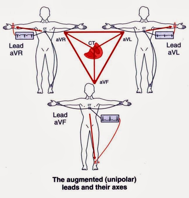
The ECG leads. Before discussing the ECG leads and various lead systems, we need to clarify the difference between ECG leads and ECG electrodes.An electrode is a conductive pad that is attached to the skin and enables the recording of electrical currents. An ECG lead is a graphical description of the electrical activity of the heart and it is created by analyzing several electrodes.