Barrett Esophagus Radiology Key
Esophagus II: Strictures, Acute syndromes, Neoplasms and Vascular impressions. Terrence C. Demos, MD, Harold V. Posniak, MD, Wayde Nagamine, MD and Mary Olson, MD. Department of Radiology of the Loyola University Medical Center, USA. In Esophagus II we will discuss: Strictures. Acute esophageal syndromes. Benign and malignant neoplasms.
Esophageal Achalasia Radiology Key
Barium esophagogram is a useful diagnostic tool for various esophageal diseases, such as strictures, diverticula, tumors, and motility disorders. This pictorial essay illustrates the typical radiographic findings of different esophageal conditions, with emphasis on the differential diagnosis and clinical implications. The article also provides some practical tips for performing and.

A barium esophagram showing a normal caliber esophagus with a large... Download Scientific Diagram
Esophagram. An esophagram is a kind of X-ray that takes video images of your esophagus in action. It's also called a barium swallow test. During the procedure, you swallow a barium contrast solution. The fluoroscopic X-ray beam visualizes your throat and esophagus while you swallow. Contents Overview Test Details.

Oesophageal atresia with tracheooesophageal fistula, ileal atresia and Hirschsprung’s disease
Esophageal varices are submucosal distal esophageal veins, connecting the portal circulation and systemic circulation, that are dilated because of portal hypertension, most commonly because of cirrhosis, resistance to portal blood flow, and increased portal venous blood inflow. Variceal rupture is the most common fatal complication of cirrhosis.

Radiographic Positioning of the Esophagus YouTube
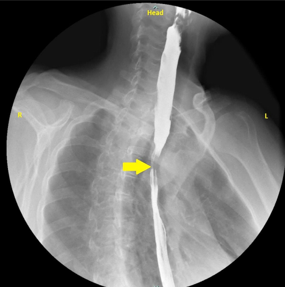
An esophageal stricture refers to the abnormal narrowing of the esophageal lumen; it often presents as dysphagia, commonly described by patients as difficulty swallowing. It is a serious sequela to many different disease processes and underlying etiologies. Its recognition and management should be prompt. Stricture formation can be due to inflammation, fibrosis, or neoplasia involving the.

Barium esophagogram before (left) and after (right) POEM in a case of... Download Scientific
∗ Department of Pediatric Gastroenterology and Nutrition, Medical University of Warsaw † Gastroenterology Unit, Bielanski Hospital, Warsaw, Poland. Address correspondence and reprint requests to Aleksandra Banaszkiewicz, MD, PhD, Department of Pediatric Gastroenterology and Nutrition, Medical University of Warsaw, Dzialdowska 1, 01-184 Warsaw, Poland (e-mail: [email protected]).

Figure. Barium esophagography showing diffuse symmetrical muscular... Download Scientific Diagram
ing this time, scrutiny of proton pump inhibitors (PPIs) has increased considerably. Although PPIs remain the medical treatment of choice for GERD, multiple publications have raised questions about adverse events, raising doubts about the safety of long-term use and increasing concern about overprescribing of PPIs. New data regarding the potential for surgical and endoscopic interventions have.

Barium swallow showing the compression in the oesophagus at the level... Download Scientific
OBJECTIVE. We evaluated the diagnostic utility of CT in emergency department (ED) patients with suspected esophageal perforation and assessed whether subsequent fluoroscopic esophagography is necessary. MATERIALS AND METHODS. This retrospective study included consecutive adult patients presenting to an urban academic tertiary care ED from January 1, 2000, to August 31, 2017, who underwent CT.
J Med Cases
Reflux esophagitis is the most common cause of esophagitis. 3 The pathophysiology leading to the development of gastroesophageal reflux is complex; however, intrinsic weakness of the lower esophageal sphincter and anatomic abnormalities, such as hiatal hernia, are likely the most common predisposing factors. 4 CT can demonstrate the previously described nonspecific signs of esophagitis as well.

Esofagograma Normal
Purpose To determine: whether the use of both esophagography (EG) and CT is superior to either study alone in the detection of esophageal injuries and perforations. Methods Paired CT and EG performed for suspected perforated or injured native esophagus (NAE) or neo-esophagus (NEOE) were retrospectively identified and independently scored for likelihood of perforation with a Likert scale.
Esofagram o bario deglutire vista laterale che mostra esofago per la diagnosi GERD o malattia da
CT Esophagography for Evaluation of Esophageal Perforation is a comprehensive review article that discusses the indications, techniques, findings, and pitfalls of this imaging modality. It provides useful tips and examples for radiologists and clinicians who encounter patients with suspected or confirmed esophageal injury. The article also compares CT esophagography with fluoroscopic.
Achalasia Esophagus Case Studies CTisus CT Scanning
Barium swallow is a dedicated test of the pharynx, esophagus, and proximal stomach , and may be performed as a single or double contrast study. The study is often "modified" to suit the history and symptoms of the individual patient, but it is often useful to evaluate the entire pathway from the lips to the gastric fundus.

Esofagograma Imagenes
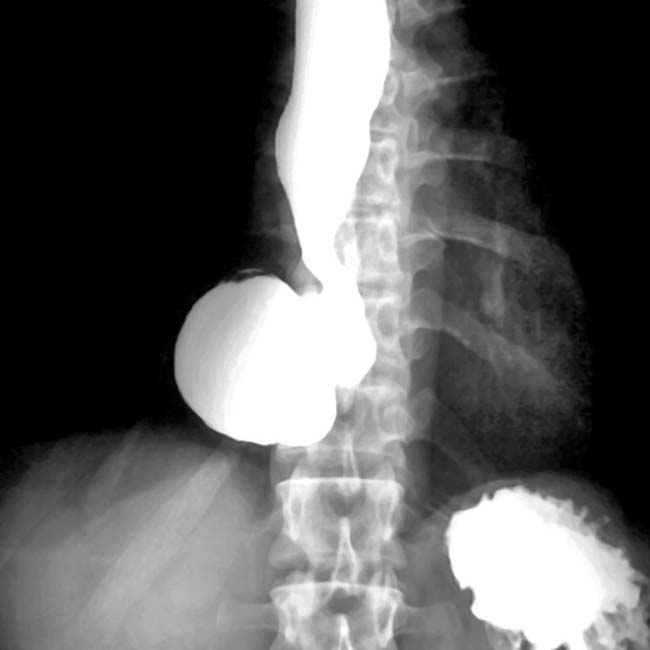
It is frequently associated with a tracheo-esophageal fistula. As such, the types of esophageal atresia / tracheo-esophageal fistula can be divided into 4: proximal atresia with distal fistula: 85%. isolated esophageal atresia: 8-9%. isolated fistula (H-type): 4-6%. double fistula with intervening atresia: 1-2%.

Esofagograma Normal
Resumen: El estudio de las enfermedades esofágicas requiere de múltiples exámenes diagnósticos, ya que ninguno, por sí solo, provee total información sobre la funcionalidad y la anatomía del tracto digestivo superior. Para los cirujanos generales y gastrointestinales, el esofagograma constituye una herramienta esencial que, además de sugerir un diagnóstico, ofrece una idea de la.
Tozande ESOFAGOGRAMMA
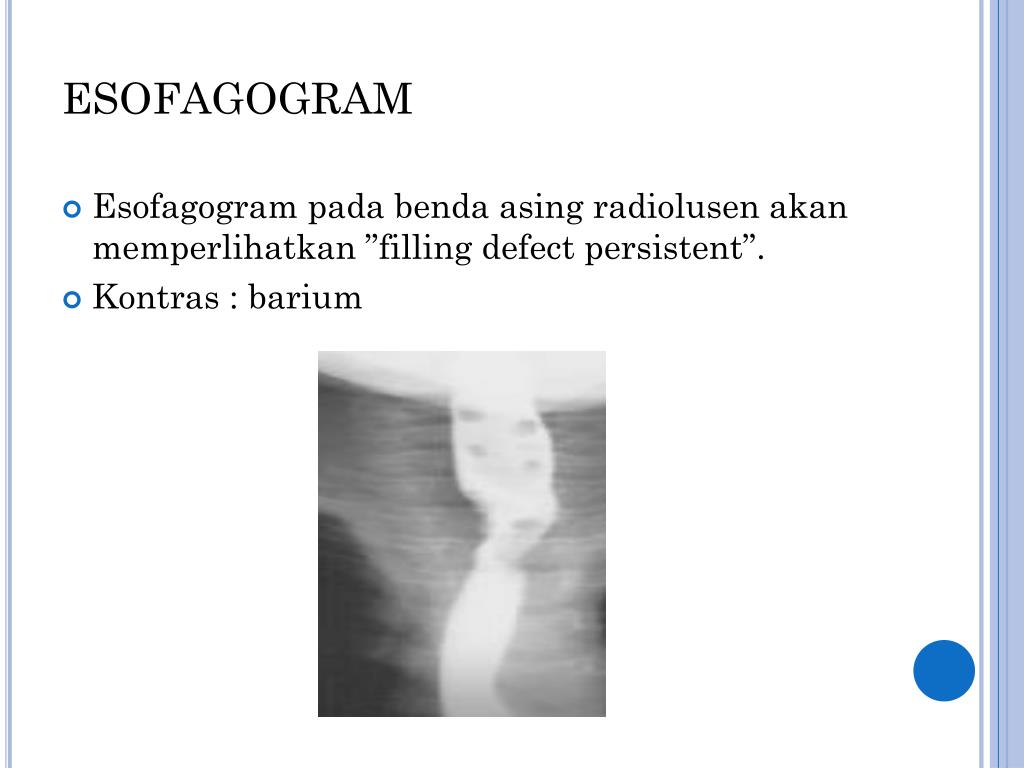
Esofagografi merupakan pemeriksaan radiologi konvensional dengan menggunakan bahan kontras yang bertujuan untuk menggambarkan lumen dari esofagus. Hasil Diterima : 3 hari Hasil Melalui : Aplikasi Halodoc (Cek riwayat buat janji) Kebijakan Pembatalan & Pengembalian Dana :
PPT dr. H. Fachzi Fitri, Sp THTKL, MAR S PowerPoint Presentation, free download ID4106045
Abstract. The barium esophagram is a valuable diagnostic test for evaluating structural and functional abnormalities of the esophagus. The study is usually performed as a multiphasic examination that includes upright double-contrast views with a high-density barium suspension, prone single-contrast views with a low-density barium suspension.