LUNGS ANATOMY wikitomy
A 62-year-old man presented with a 10-month history of dyspnea after COVID-19 infection. Dyspnea became worse in the one month preceding presentation. The chest CT showed multifocal, subpleural, bilateral opacities due to long-COVID, and infiltration around the bronchovascular bundle in the bilateral lower lung field.

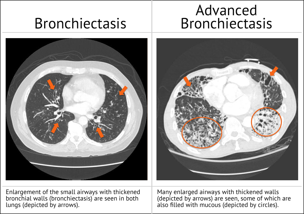
Bronchiectasis Stepwards
Double sleeve (bronchovascular) lobectomy is a feasible alternative to pneumonectomy if a complete resection can be achieved in patients with centrally located non-small cell lung cancer (NSCLC) involving both the bronchus and the pulmonary artery (PA). Traditionally, this technique has mainly been performed through a posterolateral.

Chest radiograph showing increased bronchovascular markings with right... Download Scientific
Thus, our data of bronchovascular patterns including rare anatomical variations using the accumulated 3DCTAB images of 263 patients in our institute may facilitate safe and accurate lung resection by general thoracic surgeons, regardless of the presence or absence of preoperative 3DCT images. There were several limitations in this study.

Chest Xray shows prominent bronchovascular markings
Citation, DOI, disclosures and article data. Peribronchovascular consolidation is a form of consolidation which tends to occur along peribronchovascular bundles. If smooth and mild, it may be seen as peribronchovascular thickening on chest radiography and in some instances on CT. If more extensive it tends to occur as more mass like areas of.
Image
Crowding of the bronchovascular structures is an important direct sign of volume loss and is best appreciated on contrast-enhanced CT images. The atelectatic lung enhances densely after contrast administration because of closeness of the pulmonary arteries and arterioles within the collapsed lobe.

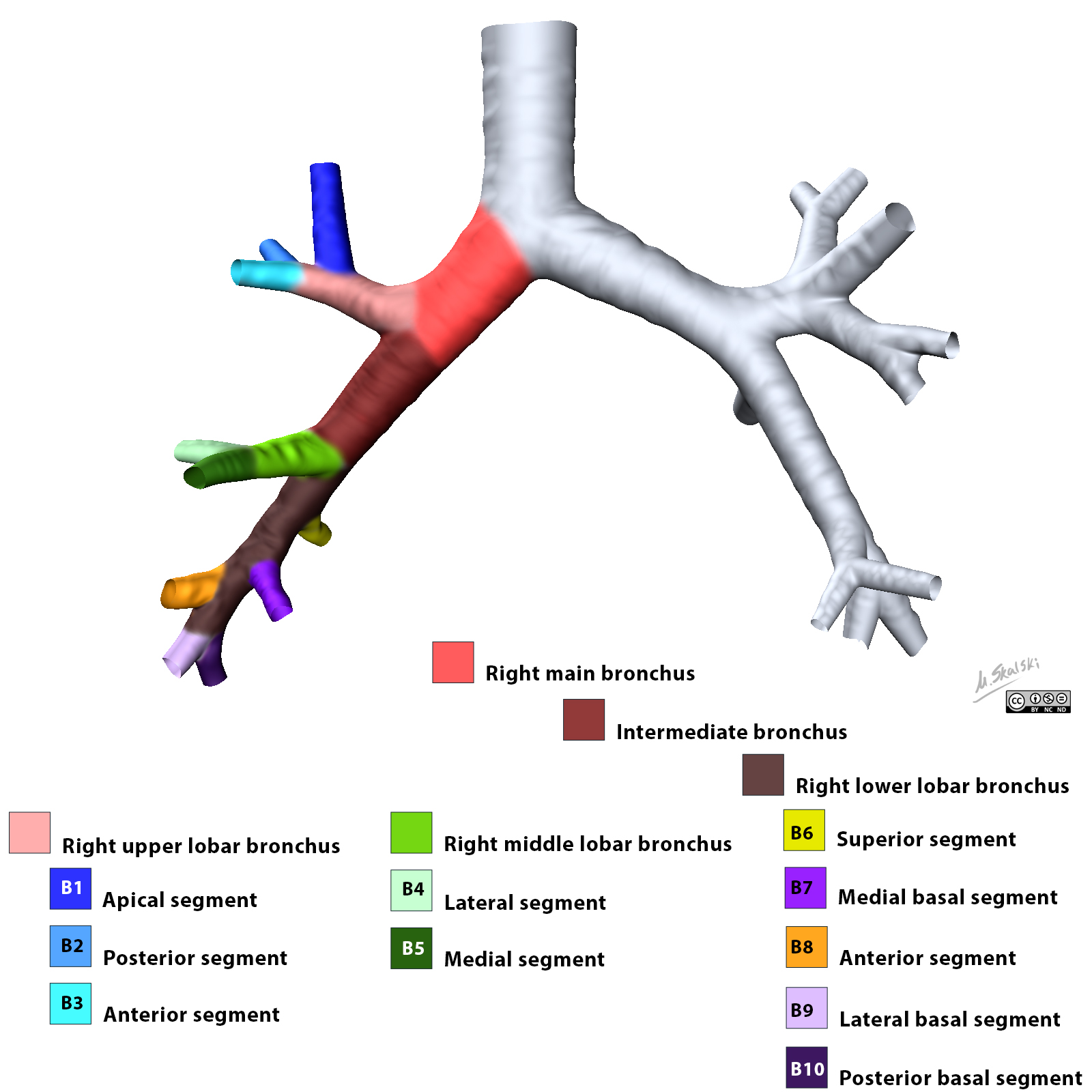
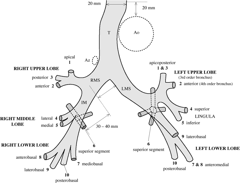
Bronchopulmonary segments annotated CT Radiology Case Bronchopulmonary
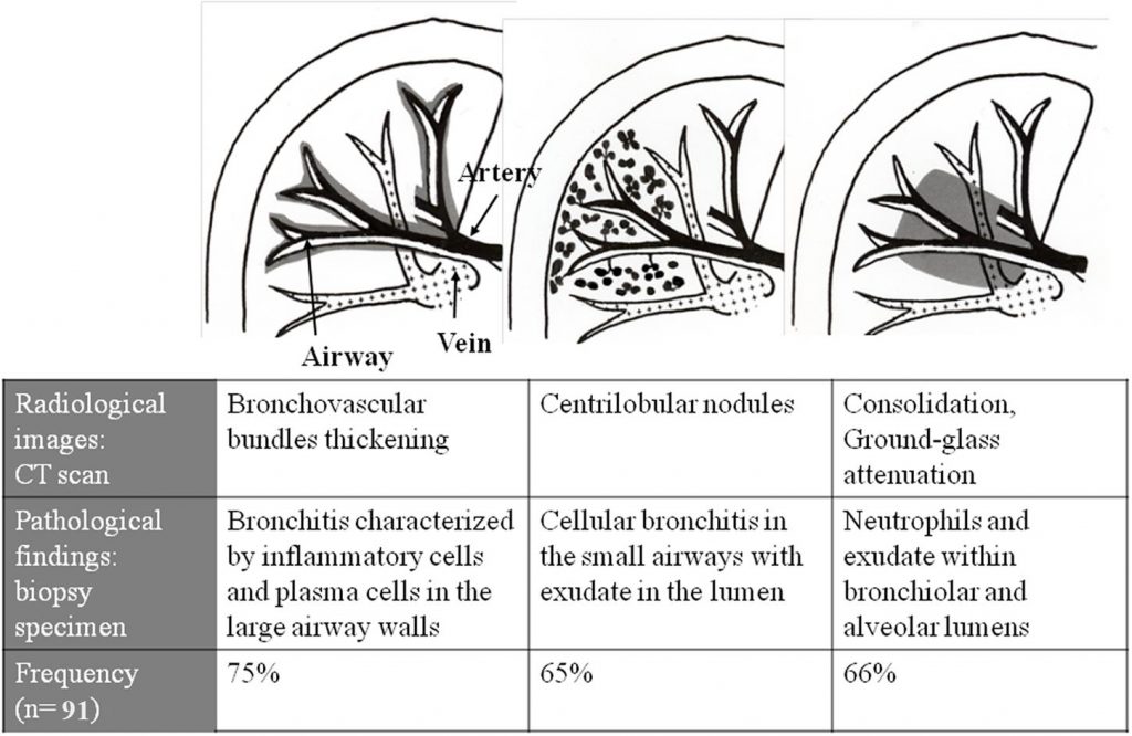
Centrilobular nodules are seen along the bronchovascular bundle (Fig 3B), indicating that the disease process is along the airway, most likely due to infection or inflammation. The nodules can be discrete or ill-defined (Fig. 15). On high-resolution CT images, it is critical to confirm absence of interlobular septal thickening in order to.
Posteroanterior chest Xray of the patient. The figure above shows the... Download Scientific
Citation, DOI, disclosures and article data. The peribronchovascular interstitium refers to the connective tissue sheath that encloses the bronchi, pulmonary arteries, and lymphatic vessels. It extends from the hilar regions through to the lung peripheries. There are many diseases that may affect the peribronchovascular interstitium.
What is Peribronchovascular Distribution on CT imaging? Medicine Specifics
Basic Interpretation. A structured approach to interpretation of HRCT involves the following questions: What is the dominant HR-pattern: reticular. nodular. high attenuation (ground-glass, consolidation) low attenuation (emphysema, cystic) Where is it located within the secondary lobule HR-pattern: centrilobular.
5. Chest Radiology Review Manual (Dahnert, Radiology Review Manual)
19:18. Halo, terimakasih atas pertanyaannya untuk Alodokter. Corakan bronkovaskular menggambarkan aliran pembuluh darah kecil di paru. Adanya peningkatan pada corakan bronkovaskular dapat menandakan adanya beberapa kemungkinan, diantaranya adalah: gagal jantung kongestif. infeksi pada paru. bronkitis kronis. asma.

Bronchiectasis Radiology imaging, Radiology, Medical mnemonics
Bronchovascular markings refer to the patterns of blood vessels and bronchi (airways) in the lungs that are visible on a radiographic image, such as a chest X-ray. These markings are normally present in the lungs and help facilitate the exchange of oxygen and carbon dioxide. Prominent bronchovascular markings occur when these patterns become.
Bronchiectasis FAQs Bronchiectasis and NTM Initiative
One can dif-ferentiate atelectasis from pneumonia by look-ing for direct and indirect signs of volume loss, including bronchovascular crowding, fissural displacement, mediastinal shift, and diaphrag-matic elevation. Detection of the air broncho-gram sign argues against the presence of a cen-tral obstructing lesion.

Prominent Bronchovascular Markings in Chest XRay Report All you need to know MyHealth
1. A postulated role for the bronchial circulation in the development of pulmonary congestion may be based on recent studies of bronchovascular control. 2. The bronchial circulation is the nutrient blood supply of the conducting airways and, therefore, plays an important role in the function of the.
Cureus Bilateral Hemopneumothorax in COVID19
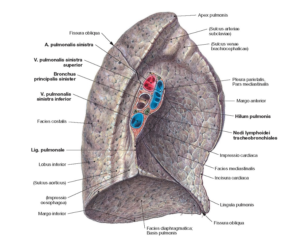
Bronchovascular bundle. Mid-size and small structures in the lung like to stick together along their path to and from the acinus, and it can generally be said that pulmonary vessels and bronchi co-occupy the space of a fibrous sheath together with a few other ancillary structures. These are described as "bronchovascular bundles".

Prominent bronchovascular structures Download Scientific Diagram
Jokoh et al., in a series of 22 patients, report CT findings comprising ill-defined centrilobular nodules in 22, ground glass opacity in 22, bronchovascular thickening in 19, subpleural nodules in 19, septal thickening in 18, cysts in 15 (1-30mm, mean 6.4mm), and lymph node enlargement in 15. 32 Large nodules on the order of 1-2cm and air-space.

(AD) Chest computed tomography showed bronchovascular bundle... Download Scientific Diagram
Understanding the meaning of prominent bronchovascular markings is crucial for patients and caregivers. It can shed light on the severity of a condition and guide further medical investigations. Significance. Prominent bronchovascular markings may suggest that there is an ongoing issue affecting the lungs or heart.

(a) Chest Xray showing prominent bronchovascular markings with basal... Download Scientific
Most common are small nodules, predominantly distributed along bronchovascular bundles, and thickened interlobular septa . Other findings include ground-glass opacities, which may be replaced by microcystic lesions, and fibrosis with honeycombing in the advanced chronic stage. Hilar or mediastinal lymphadenopathy, which always has associated.