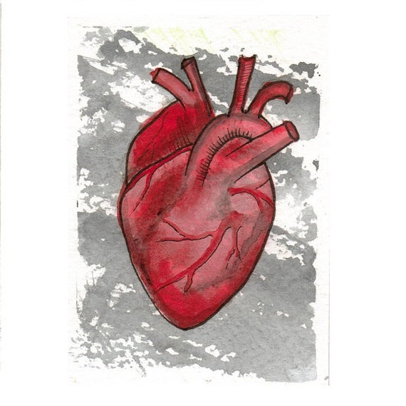
Anatomically Correct Heart Original Watercolor ACEO
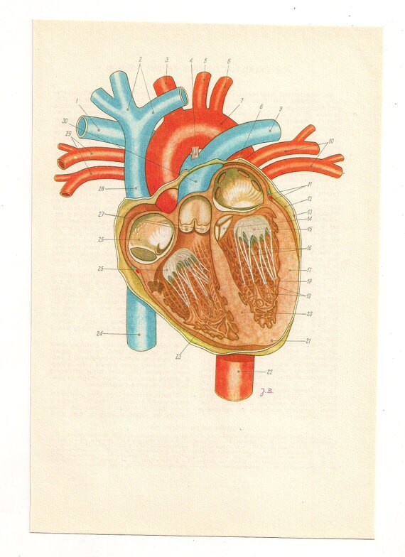
Knowing the anatomical structure of the heart is essential for understanding its function, performing clinical examination, and interpreting radiological findings. The heart consists of 4 chambers: 2 atria (ie, the right atrium and the left atrium) and 2 ventricles (ie, the right ventricle and the left ventricle).
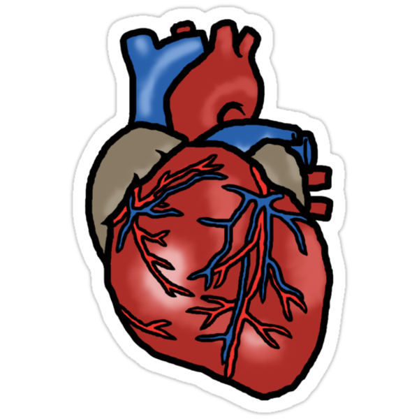
"Anatomically Correct Heart" Stickers by Bowieisgod Redbubble
Heart Wall The heart wall consists of three layers: Epicardium: The outer layer of the wall of the heart. Myocardium: The muscular middle layer of the wall of the heart. Endocardium: The inner layer of the heart. Cardiac Conduction Cardiac conduction is the rate at which the heart conducts electrical impulses.
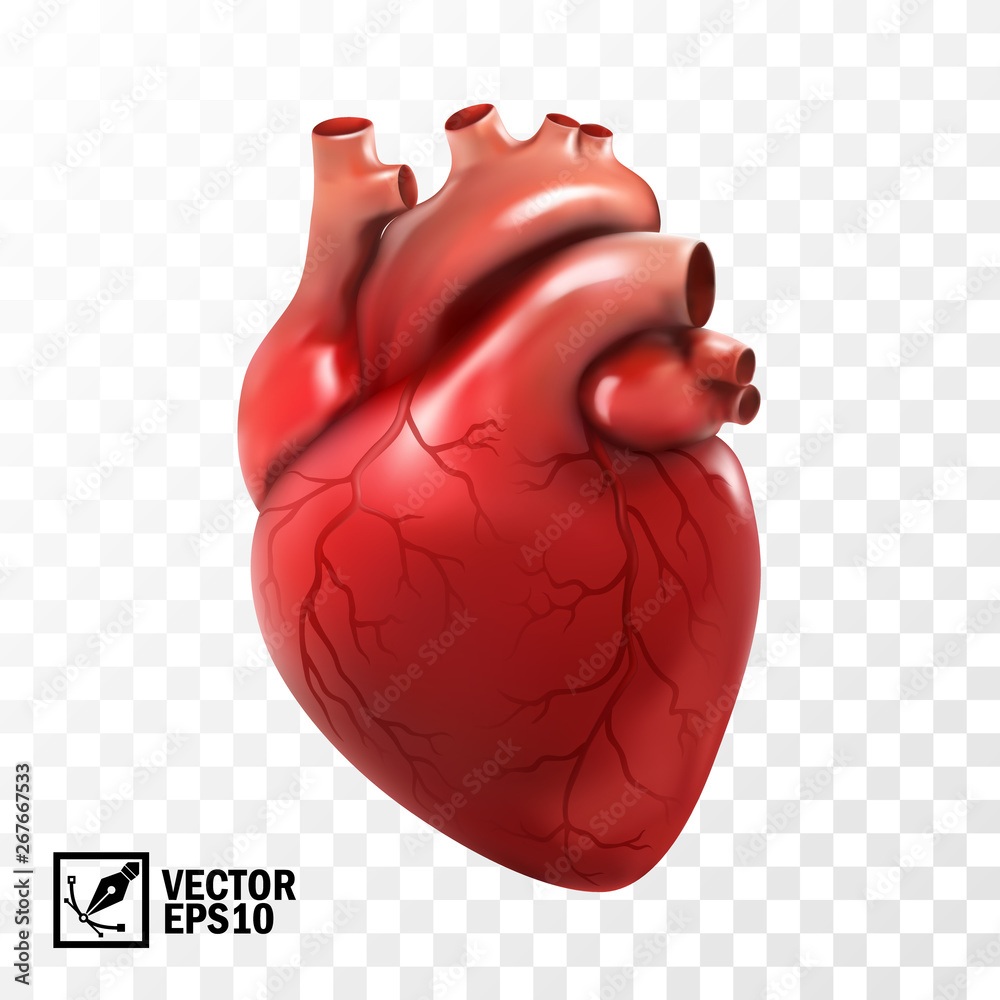
3d realistic vector isolated human heart. Anatomically correct heart
3D realistic hearts. anatomically correct hearts. Detail MRI heart. Dog MRI heart. Rabbit MRI heart. Horse MRI heart. heart structure 3D interactive virtual heart Heart : A powerful muscle slightly larger than a clenched fist. It is composed of four chambers, two upper (the atria) and two lower (the ventricles). It works as a pump to send.
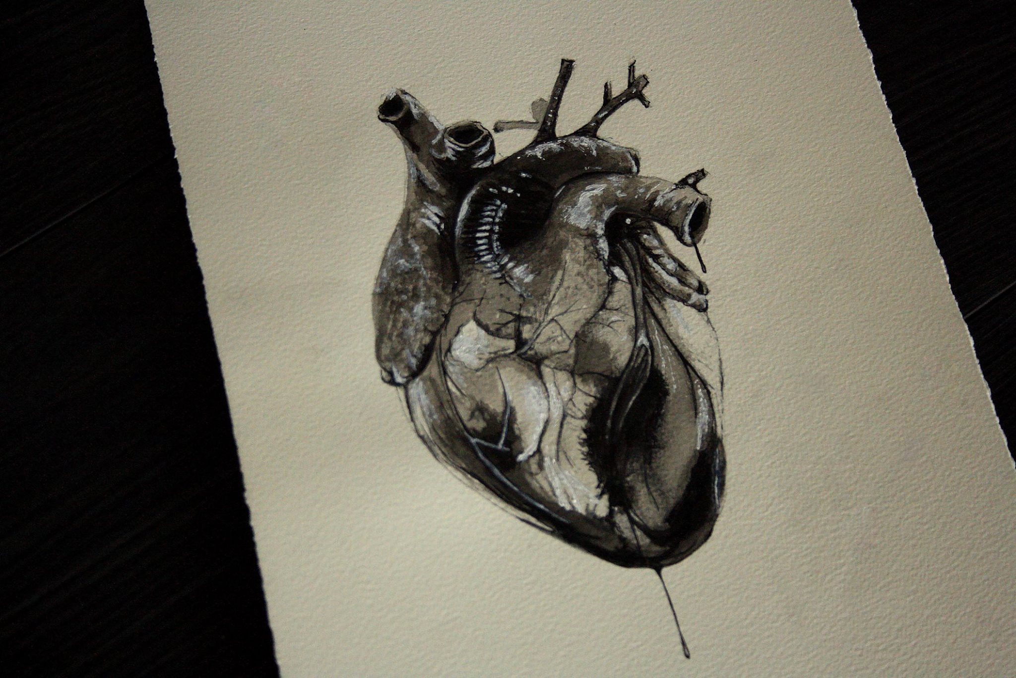
Anatomically Correct Heart Drawing at GetDrawings Free download
Heart Anatomy of the heart: anatomical illustrations and structures, 3D model and photographs of dissection Antoine MICHEAU, MD , Denis HOA, MD Authors affiliations Publication date: Aug 8, 2008 | Last update: Oct 3, 2022 https://doi.org/10.37019/e-anatomy/180 ISSN 2534-5079

Anatomical Heart Drawing Clipart Michael Arntz
The human heart is located within the thoracic cavity, medially between the lungs in the space known as the mediastinum. Figure 19.2 shows the position of the heart within the thoracic cavity.
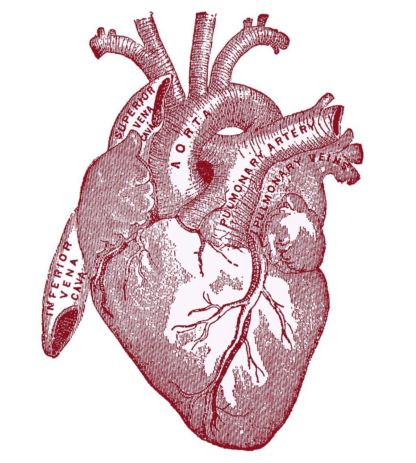
8 Anatomical Heart Drawings! The Graphics Fairy
The heart has four chambers: the left and right atriums and the left and right ventricles. They form a shallow groove at the line of their junction, which form the atrioventricular groove. The atrioventricular groove hosts major coronary arteries while they travel along to the line of attachment of atrioventricular valves.
Anatomically correct heart Anatomical anatomy art poster
Anatomically Correct Heart (1 - 60 of 360 results) Price ($) Shipping All Sellers Anatomical Heart Necklace, anatomically correct heart jewelry, realistic human heart necklace, I love you necklace, meaningful necklace gift (1.2k) $12.95

Anatomically Correct Heart Drawing Free download on ClipArtMag
The heart holds a special position in anatomical sciences. For instance, you can live without your spleen or with only one kidney, you can even regrow your liver-but you cannot live without a heart. This page will introduce you to the anatomy of the heart. Contents Heart anatomy Heart valves Blood flow through the heart Coronary circulation

Dotwork Anatomically Correct Heart and Arrow 15x7cm Printed on 300gsm
Heart Anatomy Your heart is located between your lungs in the middle of your chest, behind and slightly to the left of your breastbone (sternum). A double-layered membrane called the pericardium surrounds your heart like a sac.

Anatomically Correct Heart Drawing at GetDrawings Free download
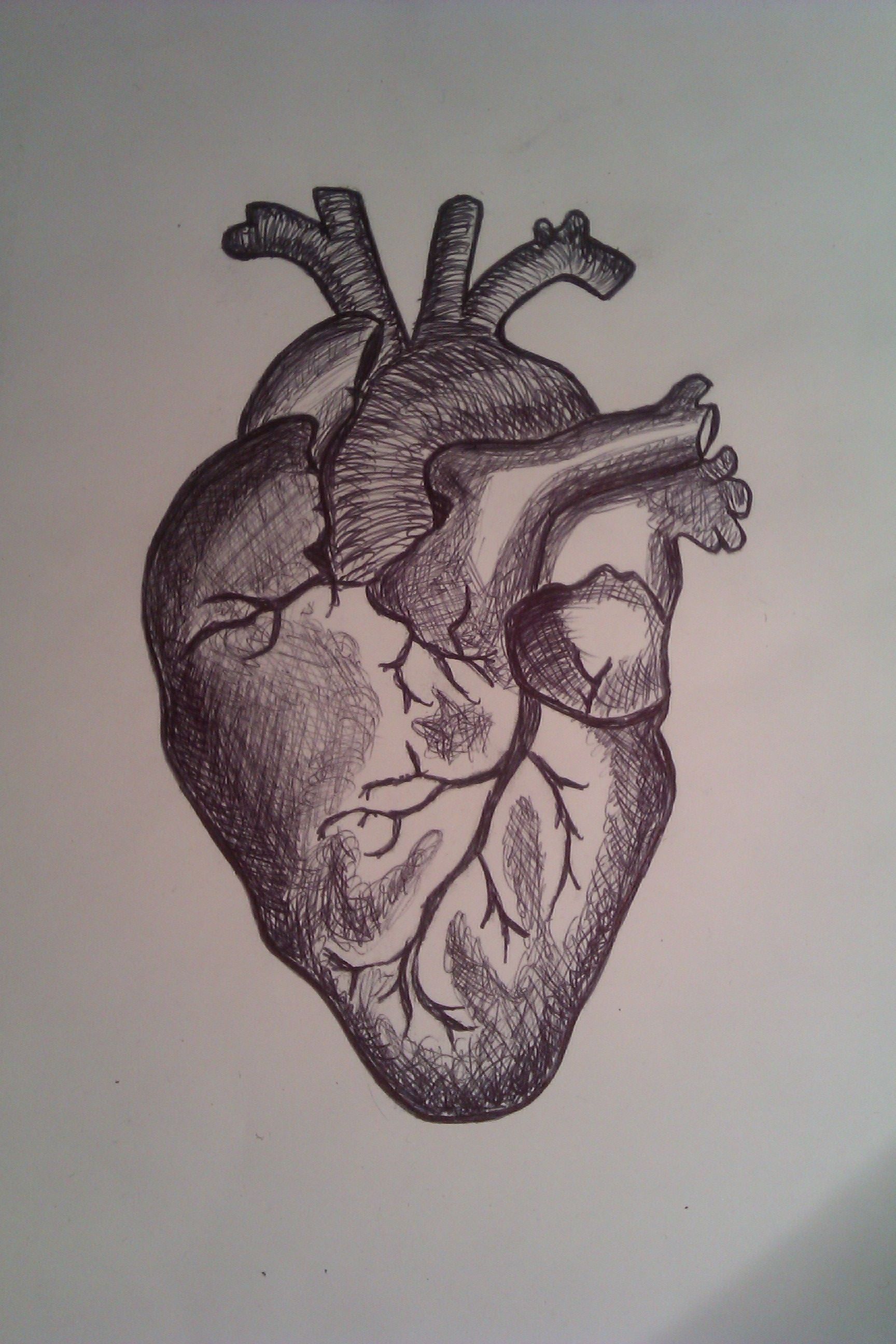
1 Draw a tilted and irregular curved shape in the center of your page. Use a pen or pencil to draw the heart's main body. Create a curved shape similar to an acorn or apple's bottom half. Angle the slightly tampered end of the shape to the left about 120 degrees. [1] The main shape will be the basis for the left and right ventricles.

How To Draw An Anatomically Correct Heart at How To Draw
Many physicians and medical students can envision an anatomically correct heart, in addition to the anatomical conformation of congenital heart defects.

Anatomically Correct Heart Drawing at GetDrawings Free download
A human heart viewed from the so-called anterior position, demonstrating the valentine heart orientation used by many to incorrectly describe anatomy. The red line surrounding the heart is the characteristic symbol, which was theoretically derived from observing the heart in this orientation.
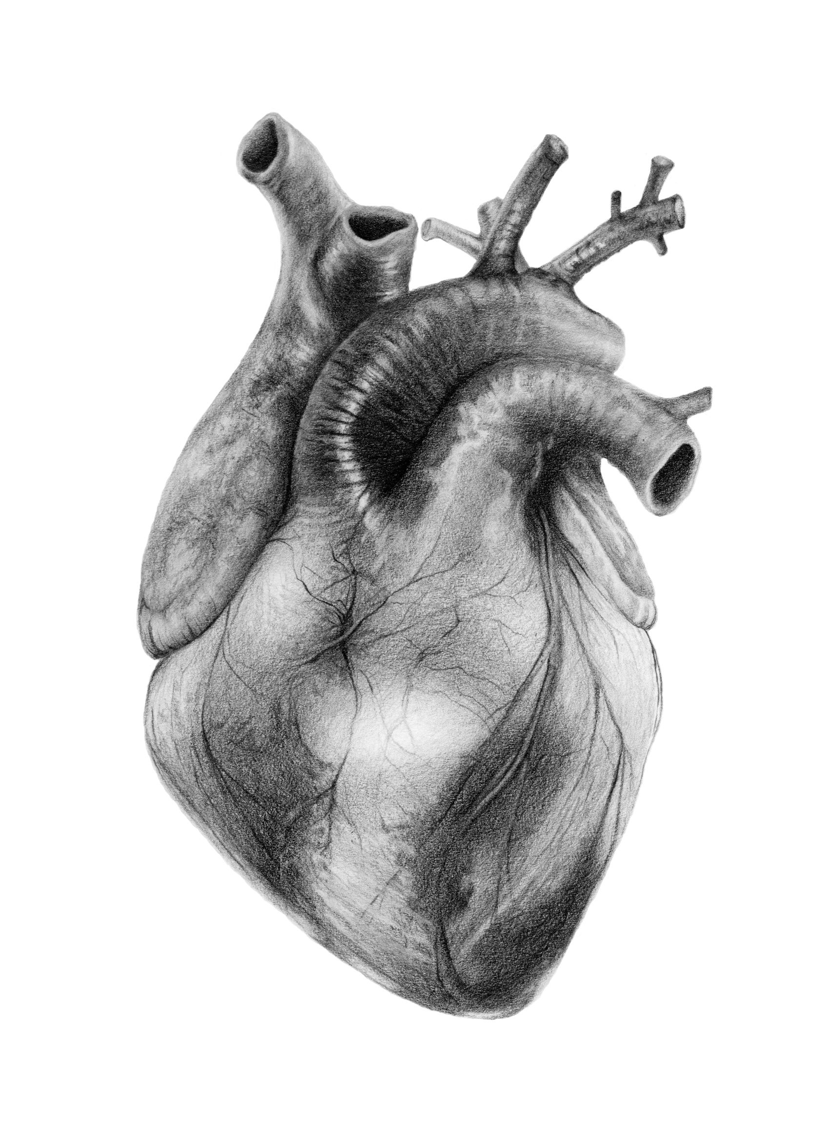
Anatomically Correct Heart Drawing at Explore
The anatomical base points dorsally and slightly upwards to the right. The right atrium and ventricle make up most of the sternocostal surface of the heart, while the diaphragmatic surface is made up by the right and left ventricles. The left atrium contributes to the anatomical base. Endocardium. 1/3.
Anatomically Correct Heart Drawing at GetDrawings Free download
Heart conditions are among the most common types of disorders affecting people. In the United States, heart disease is the leading cause of death for people of all genders and most ethnic and racial groups. Common conditions that affect your heart include: Atrial fibrillation (Afib): Irregular electrical impulses in your atrium.

Anatomically Correct Human Heart Diagram
PLACING THE HEART IN THE CHEST. One of the first rules of human anatomy is that all bodily parts should be described as viewed in the so‐called anatomical position (Anderson and Loukas, 2009).This means that the heart should be described as it is normally positioned within the thorax (Fig. (Fig.1; 1; Cosío et al., 1999; Anderson et al., 2013).It is difficult in the dissecting room, however.
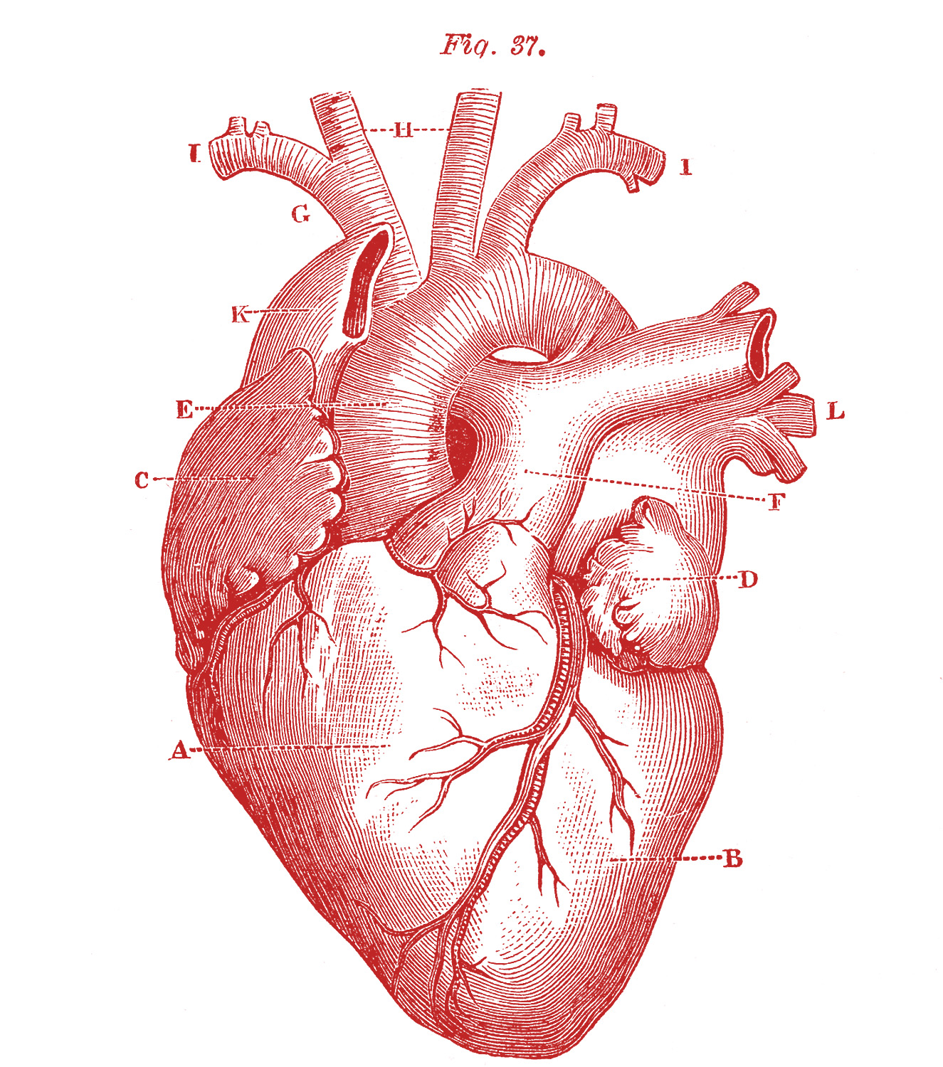
Royalty Free Images Anatomical Heart Vintage The Graphics Fairy
The heart is an organ that weighs approximately 350 grams (less than one pound). It's nearly the size of an adult's clenched fist. It's located in the thorax (chest)—between the lungs —and extends downward between the second and fifth intercostal (between the ribs).