
3D Ultrasound at 16 Weeks — The Bump
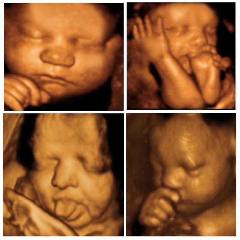
A 3D ultrasound of a human fetus aged 20 weeks. 3D ultrasound is a medical ultrasound technique, often used in fetal, cardiac, trans-rectal and intra-vascular applications. 3D ultrasound refers specifically to the volume rendering of ultrasound data. When involving a series of 3D volumes collected over time, it can also be referred to as 4D ultrasound (three spatial dimensions plus one time.
16 week 3D ultrasound boy or girl?! Help?!
When is the Best Time to Get a 3D Ultrasound? by uipadmin | Nov 6, 2015 | Uncategorized Owning an elective ultrasound facility, I often get the age old question "When is the best time to come in for a 3D/4D ultrasound?" My facetious response: When you're pregnant. My professional business response: Well, that depends…
The Baby Scene Perth
A 3D/4D Ultrasound taken of a baby AT 16 weeks that visited Prenatal Impressions, the only premier provider of 3D/4D Ultrasounds in Orlando, Florida since 20.

3D ultrasound 16 weeks its a boy! YouTube
Transvaginal ultrasound also makes it easier to diagnose early pregnancy problems, such as a miscarriage or a molar or ectopic pregnancy. Not all women have a first-trimester ultrasound. It's standard to have just one ultrasound during pregnancy - a mid-pregnancy transabdominal ultrasound between 18 and 22 weeks.

4D ultrasound Baby Scan 16 weeks pregnant ultrasound, Baby scan, 16
What is a 3D or 4D ultrasound? Keepsake ultrasound pictures and videos are popular, but many healthcare providers advise against them. Here's why. Medically reviewed by Cheryl Axelrod, M.D., ob-gyn Written by Deepi Brar | Mar 3, 2021 Photo credit: iStock What are 2D, 3D, and 4D ultrasounds? What ultrasounds will I have during pregnancy?
Smithback Twins 16 Week Ultrasound Pictures!
Like their two-dimensional (2D) counterparts, 3D ultrasounds use high-frequency sound waves and special imaging software to create images of your baby's soft tissues, organs, and other anatomy..
3D & 4D Ultrasound 16 weeks YouTube
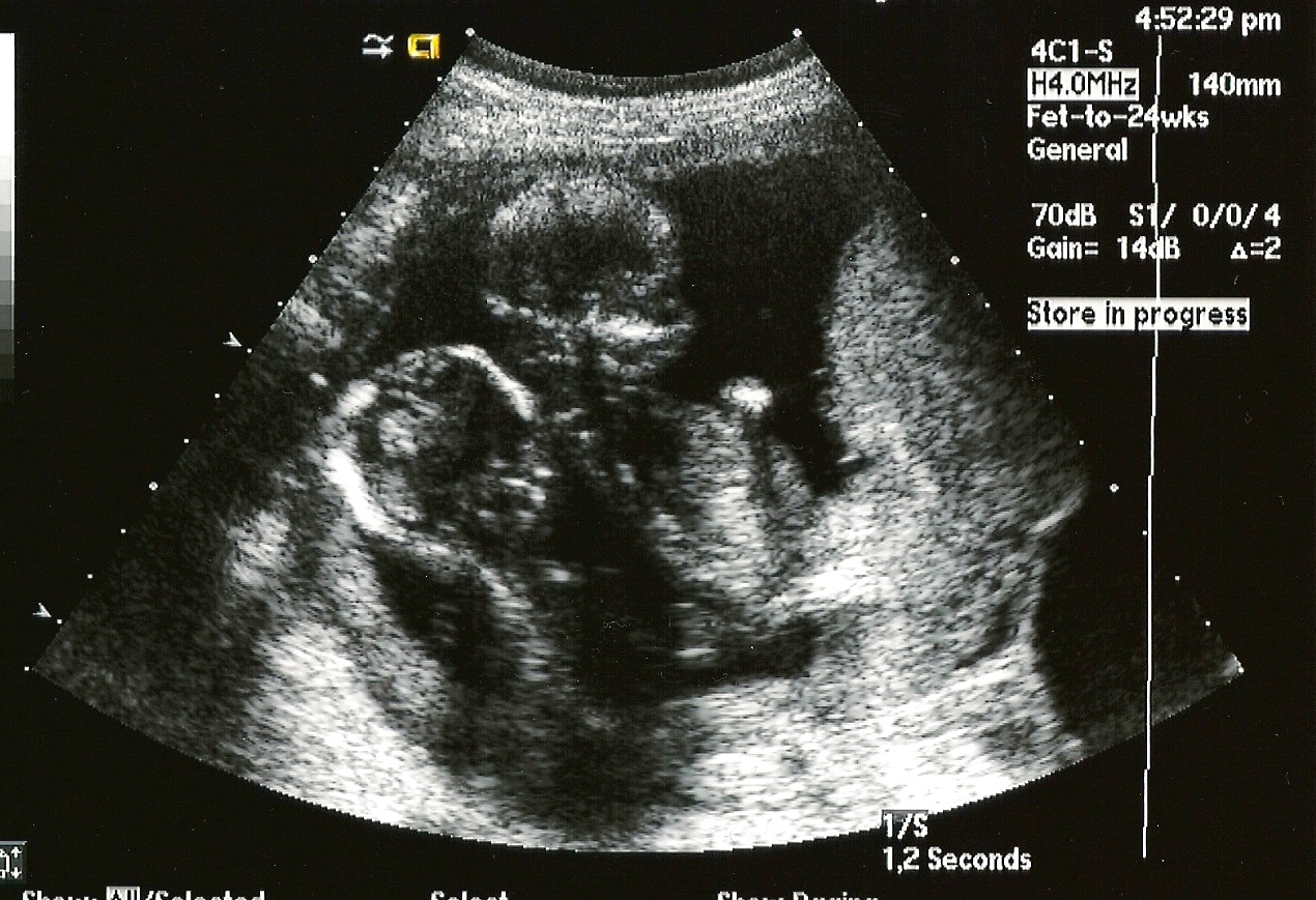
Fetal ultrasound is used to check that the heart is working properly and to see if there could be any heart problems. The brain Below is an image of the base of the brain, called the cerebellum. This type of image usually is taken during an ultrasound done between 18 and 22 weeks of pregnancy.

3D ultrasound testimonials 3D & 4D Ultrasound in Ft. Myers, FL
Like regular ultrasounds, 3D and 4D ultrasounds use sound waves to create an image of your baby in your womb. What's different is that 3D ultrasounds create a three-dimensional image of your baby.

16 Weeks Pregnant Ultrasound
3.5 ounces Baby is as big as an avocado save article By The Bump Editors | Updated March 30, 2023 Medically Reviewed by Patricia Pollio, MD In this article: Key takeaways at week 16 Video recap at 16 weeks Baby's development at week 16 3D anatomy views Pregnancy symptoms this week Your body at 16 weeks Tips for week 16 Checklist for week 16

3D Ultrasound at 16 weeks pregnant During the 16th week of pregnancy
A 16-week ultrasound isn't standard — but don't let it worry you! Look at it as an opportunity to get more glimpses of baby. Your first ultrasound will usually occur between 8 and 14.
.jpg)
[最新] 24 weeks pregnant ultrasound 3d 214767Can you get a 3d ultrasound
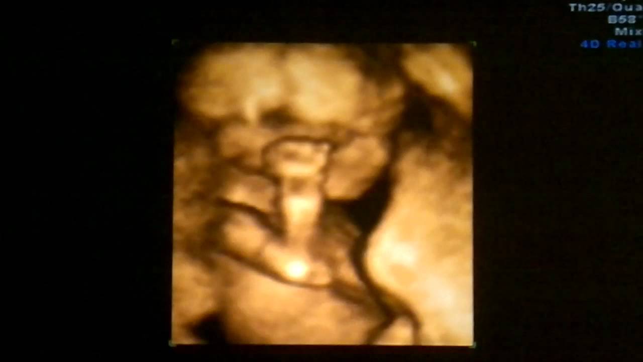
27 Comments. I did one at 19 weeks and my boy totally looked like an alien to me lol. Did one again at 32 weeks and he looked like a beautiful baby. Here's the side by side. At 16 weeks they likely will look like an alien but you can still do the ultrasound!!

Baby Heise 16 week 3d ultrasound baby Heise 3d ultrasound … Flickr
Clearer Visualization. One of the best things about a 27-week 3D ultrasound is that it takes clear pictures. Imagine looking at a very detailed photo. That's what a 3D ultrasound does for images.

22 week 3D Ultrasound Pictures A Date With Baby
16 WEEK GENDER REVEAL! AMAZING 3D & 4D ULTRASOUND! SparklingReview 7.99K subscribers Subscribe Subscribed 1.1K Share 590K views 9 years ago It's A We're so excited to meet our little one!.

3d Baby Ultrasound Edmonton Get More Anythink's
Pregnancy checklist at 16 weeks pregnant Investigate second-trimester prenatal tests. New trimester, new prenatal tests.Around 16 to 18 weeks, you may be offered a test for Alpha Fetal Protein (AFP) to help screen for neural tube defects (problems with the brain and spinal cord), such as spina bifida. (This test isn't as accurate as the anatomy ultrasound, however, which you'll have in a few.

Pin on Olivia Rian 2018
Furthermore, at A Date With Baby 4D Ultrasound, owing to our superior technology and trained sonographers, we can determine the gender a bit earlier, starting at around 15-16 weeks. At the 18-week ultrasound, the doctor will look at how the baby is growing and check on the health of the pregnancy. This will involve a detailed scan of the baby.

This is our 4d baby scan done our clinic Baby Moments. Look at the baby
Your baby at 16 weeks' ultrasound is the largest it has been in your pregnancy till date, and few things need to be done before you go for a 16-week scan. Your baby which measures around four and a half inches during the 16 th week will grow double in size very quickly, here onwards, month on month.