
The Structure Of Human Skin Cells Stock Illustration Download Image Now Hair Follicle
Stratum basale, also known as stratum germinativum, is the deepest layer, separated from the dermis by the basement membrane (basal lamina) and attached to the basement membrane by hemidesmosomes. The cells found in this layer are cuboidal to columnar mitotically active stem cells that are constantly producing keratinocytes.
Schematic representation of skin structure and cell population. The... Download Scientific Diagram
Dermatology nomenclature Desquamation imbalance Psoriasis Albinism Sources + Show all Without the skin, humans would be susceptible to a myriad of pathologies. The organ acts as a protective barrier that limits the migration of microbes and chemicals into the body.
Skin diagram to label Labelled diagram
Facts about the skin. The skin is the body's largest organ. It covers the entire body. It serves as a protective shield against heat, light, injury, and infection. The skin also: Regulates body temperature. Stores water and fat. Is a sensory organ. Prevents water loss. Prevents entry of bacteria. Acts as a barrier between the organism and its.

Anatomy of human skin. The most superficial layer of the skin is the... Download Scientific
The skin is the body's largest organ, made of water, protein, fats and minerals. Your skin protects your body from germs and regulates body temperature. Nerves in the skin help you feel sensations like hot and cold. Your skin, along with your hair, nails, oil glands and sweat glands, is part of the integumentary (in-TEG-you-ME I NT-a-ree) system.

Structure Of Skin Skin Structure and Function LearnFatafat
Label Me! Printout The skin is an organ that forms a protective barrier against germs (and other organisms) and keeps the inside of your body inside your body, and keeps what's outside of your body outside. Skin also helps maintain a constant body temperature. Human skin is only about 0.07 inches (2 mm) thick.

Skin Definition, Structure And Functions Of Skin
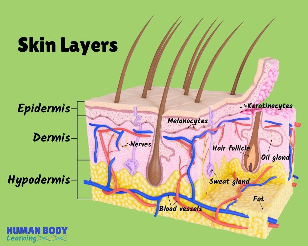
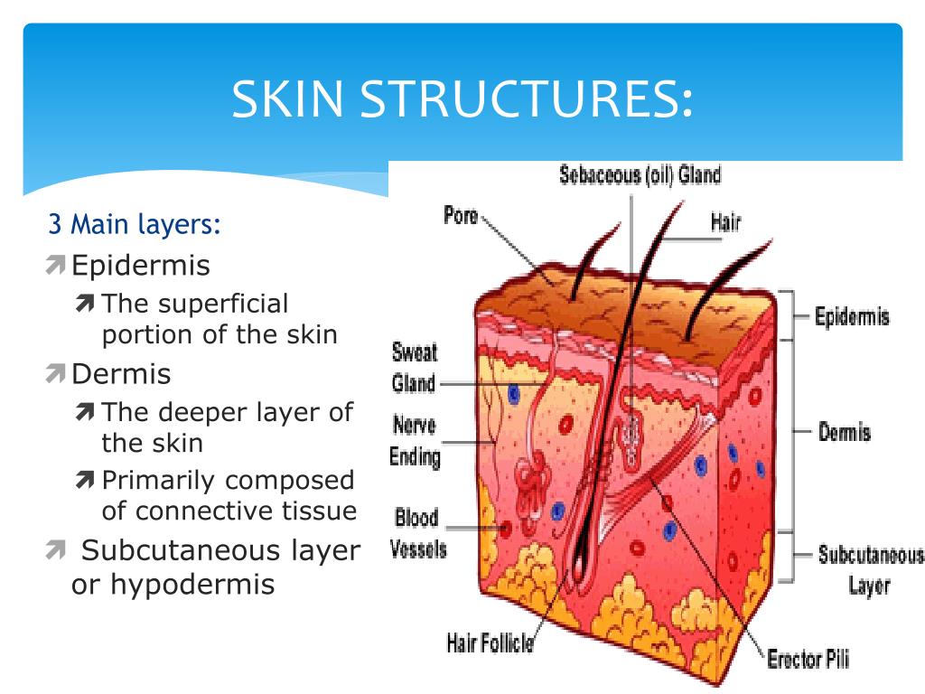
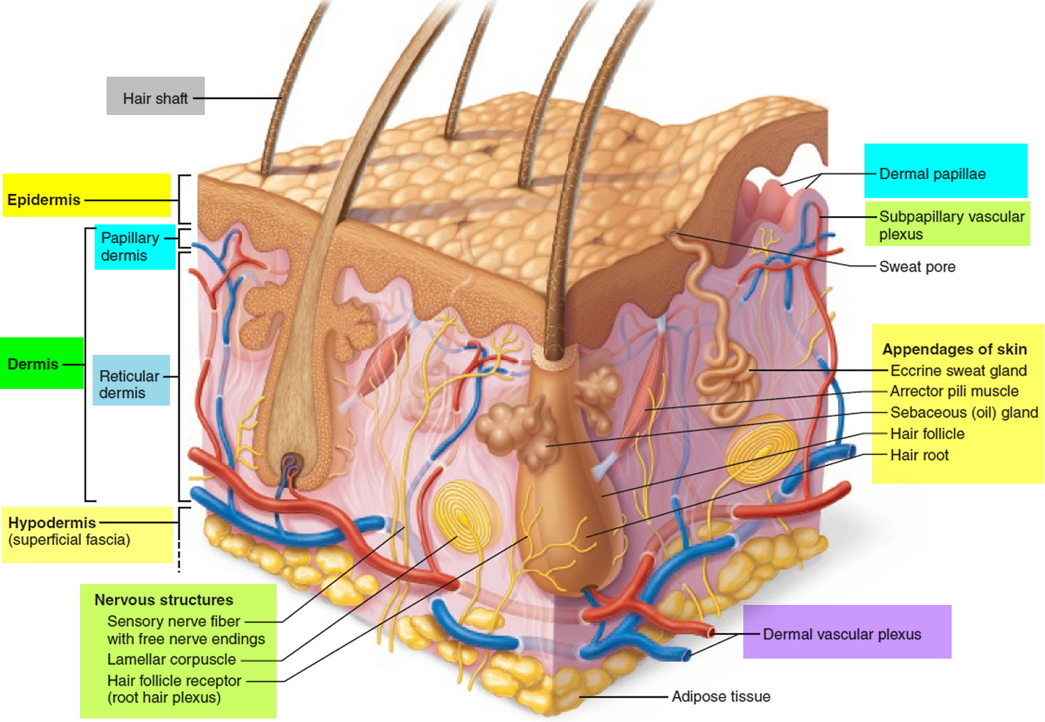
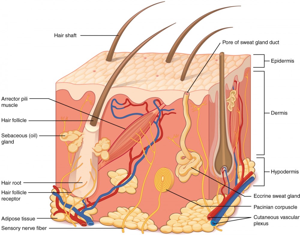
The Dermis Hypodermis The number of skin layers that exists depends on how you count them. You have three main layers of skin—the epidermis , dermis, and hypodermis (subcutaneous tissue). Within these layers are additional layers. If you count the layers within the layers, the skin has eight or even 10 layers.
The skin Understanding cancer Macmillan Cancer Support
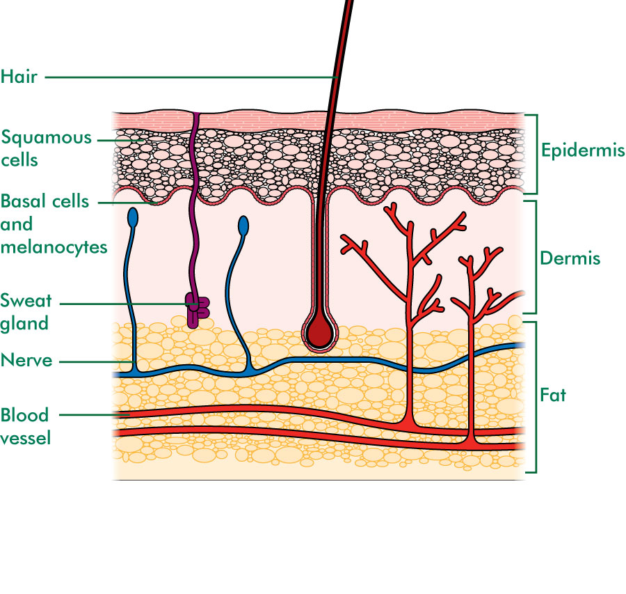
Above: Skin cross section diagram showing the epidermis with melanocytes (pigment cells), dermis, and hypodermis (fatty tissue) layers as well as glands, hairs, and blood vessels embedded in these tissue layers. The skin is also called the cutaneous membrane. There are two types of skin: thin skin that is covered with hair (also contains.

Human skin diagram Subcutaneous tissue, Skin structure, Epidermis
This diagram shows the layers found in skin. There are three main layers: the epidermis, dermis and hypodermis. There are also sweat glands, and hairs, which have sebaceous glands, and a smooth muscle called the arrector pili muscle, associated with them.
Skin Layers Anatomy Diagram for Kids
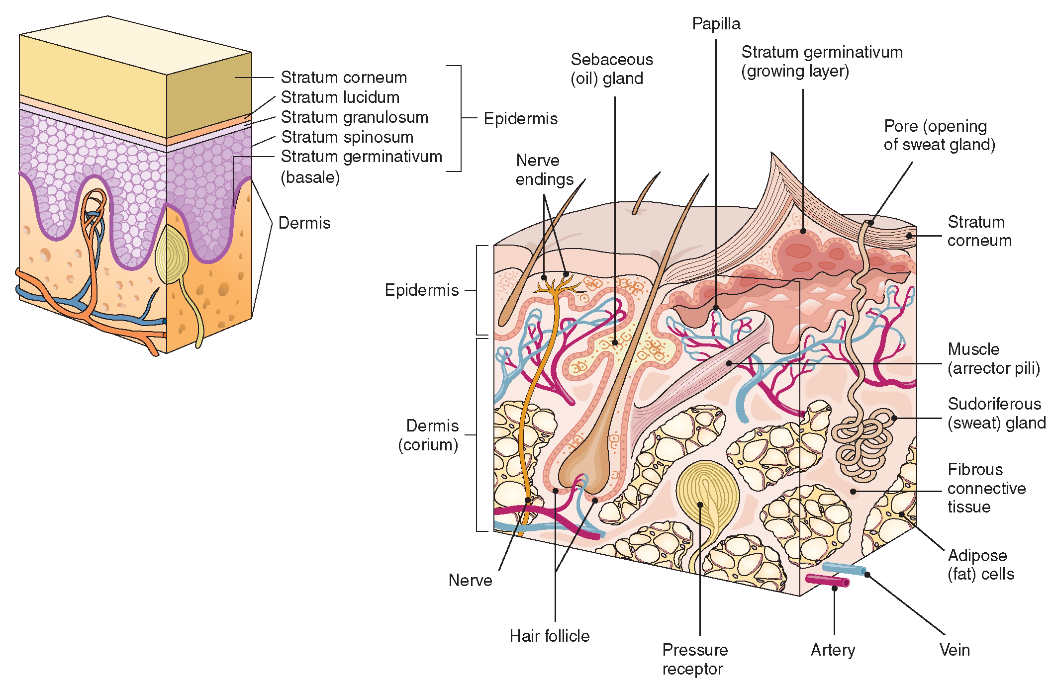
Figure 1. Layers of Skin. The skin is composed of two main layers: the epidermis, made of closely packed epithelial cells, and the dermis, made of dense, irregular connective tissue that houses blood vessels, hair follicles, sweat glands, and other structures.

The structure of the skin is composed of two layers (1) the epidermis... Download Scientific
Reading time: 16 minutes Recommended video: Integumentary system [19:28] Structure and layers of the skin. Thick skin 1/10 Synonyms: none The integumentary system is the body system which surrounds you, both literally and metaphorically speaking.
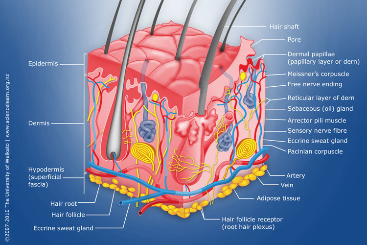
Diagram of human skin structure — Science Learning Hub
Explore Skin Diagram with BYJU'S. Diagram of the skin is illustrated in detail with neat and clear labelling. Also available for free download
PPT Basic Skin Structure PowerPoint Presentation, free download ID6099891
Dermis. Definition. Fibrous and elastic tissue, provides strength and elasticity to the skin and supports the epidermis, home to hair follicles, glands, nerves etc. Location. Term. Papillary Layer. Definition. Upper dermal layer, provides the epidermis with nutrients and regulates body temperature. Location.
The Integumentary System (Structure and Function) (Nursing) Part 1
Figure 1. The skin is composed of two main layers: the epidermis, made of closely packed epithelial cells, and the dermis, made of dense, irregular connective tissue that houses blood vessels, hair follicles, sweat glands, and other structures. Beneath the dermis lies the hypodermis, which is composed mainly of loose connective and fatty tissues.

Skin Structure infographic LifeMap Discovery
The Epidermis The epidermis is composed of keratinized, stratified squamous epithelium. It is made of four or five layers of epithelial cells, depending on its location in the body. It does not have any blood vessels within it (i.e., it is avascular). Skin that has four layers of cells is referred to as "thin skin."
Dermis Layers, Papillary Layer, Function Epidermis
Figure 5.1.1 - Layers of Skin: The skin is composed of two main layers: the epidermis, made of closely packed epithelial cells, and the dermis, made of dense, irregular connective tissue that houses blood vessels, hair follicles, sweat glands, and other structures.
Layers of the Skin Anatomy and Physiology I
The epidermis is the thin outer layer of the skin. It consists of 2 primary types of cells: Keratinocytes. Keratinocytes comprise about 90% of the epidermis and are responsible for its structure and barrier functions. Melanocytes. Melanocytes are found at the base of the epidermis and make melanin. This gives the skin its color. Dermis