Understanding how the ear works Hearing Link Services
The ear canal, or auditory canal, is a tube that runs from the outer ear to the eardrum. The ear has outer, middle, and inner portions. The ear canal and outer cartilage of the ear make.
Your Hearing Heritage Hearing
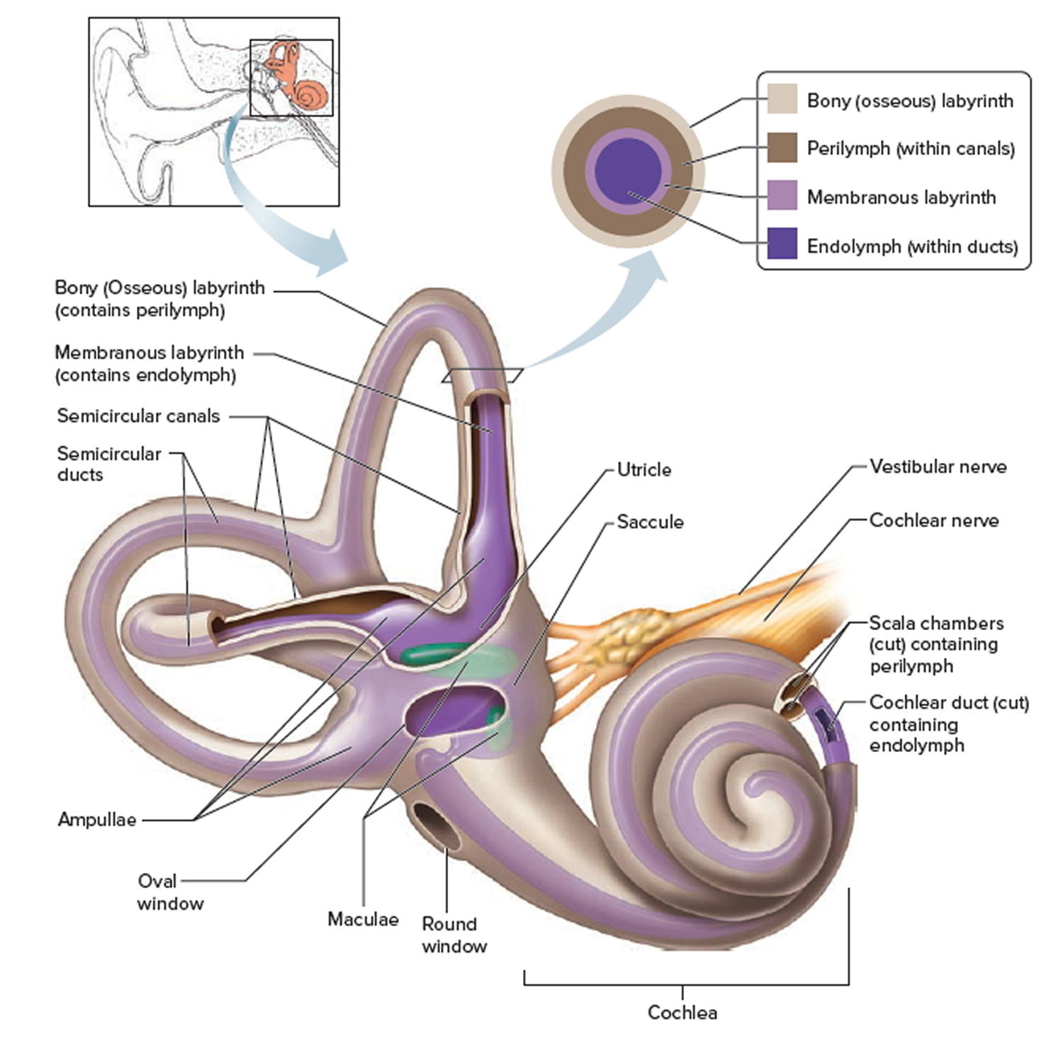
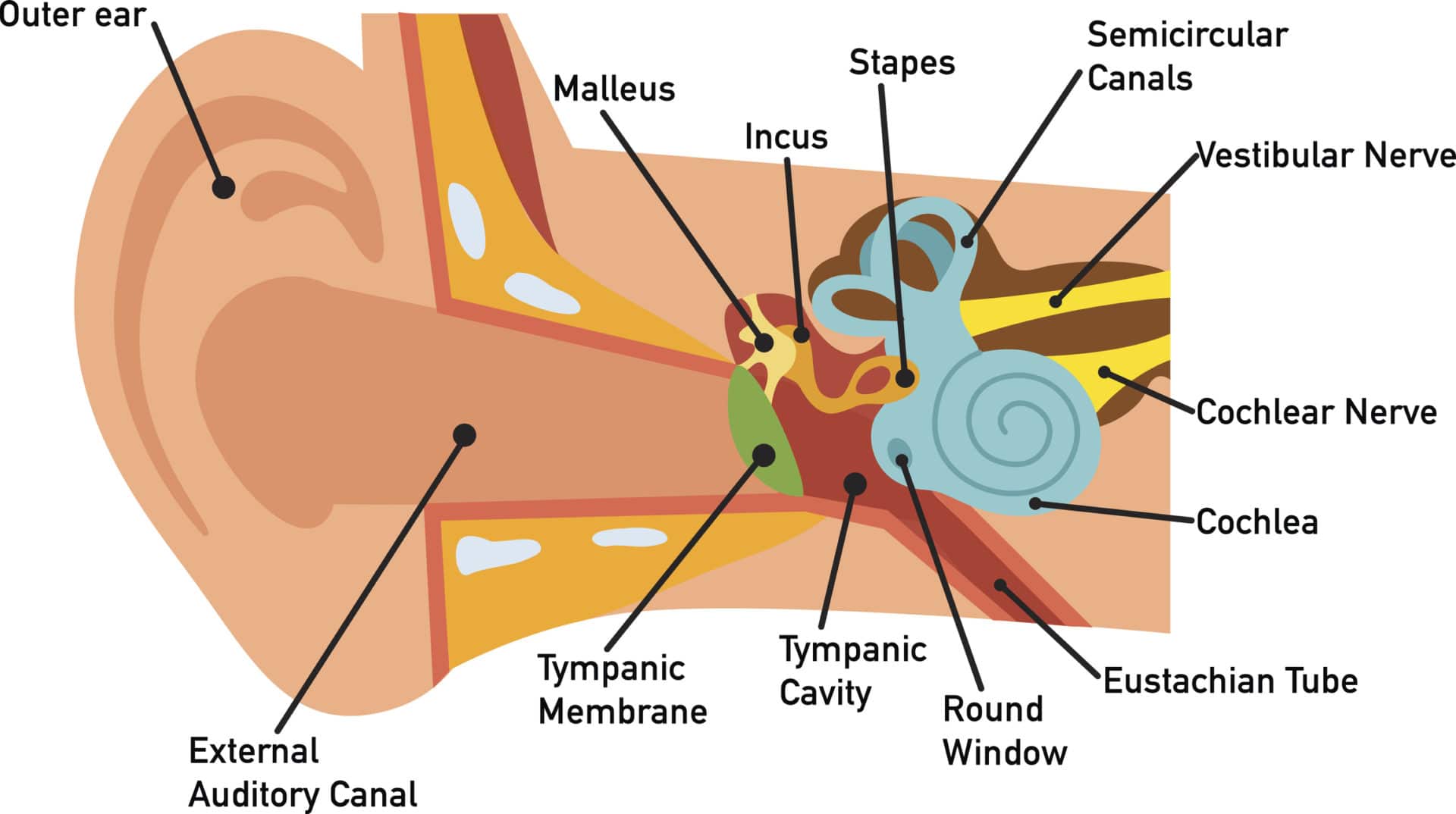
The inner ear has two openings into the middle ear, both covered by membranes. The oval window lies between the middle ear and the vestibule, whilst the round window separates the middle ear from the scala tympani (part of the cochlear duct). Bony Labyrinth. The bony labyrinth is a series of bony cavities within the petrous part of the temporal.
Discovering Something New ongoing learning How the ear works
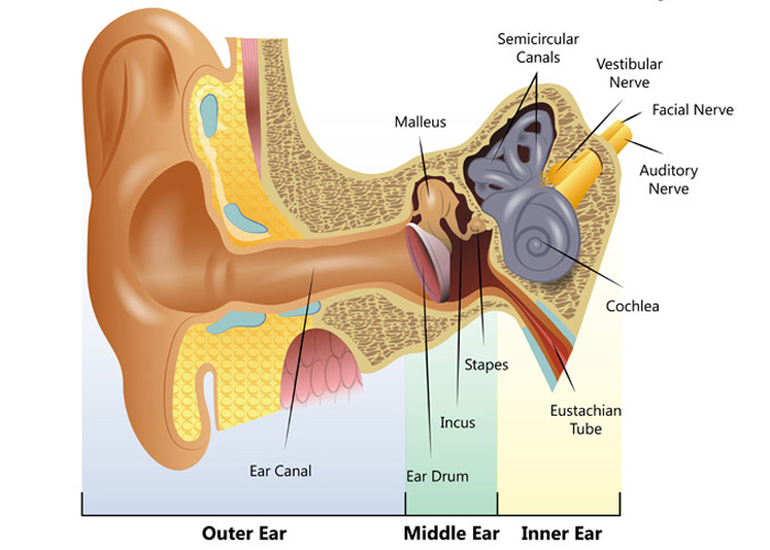
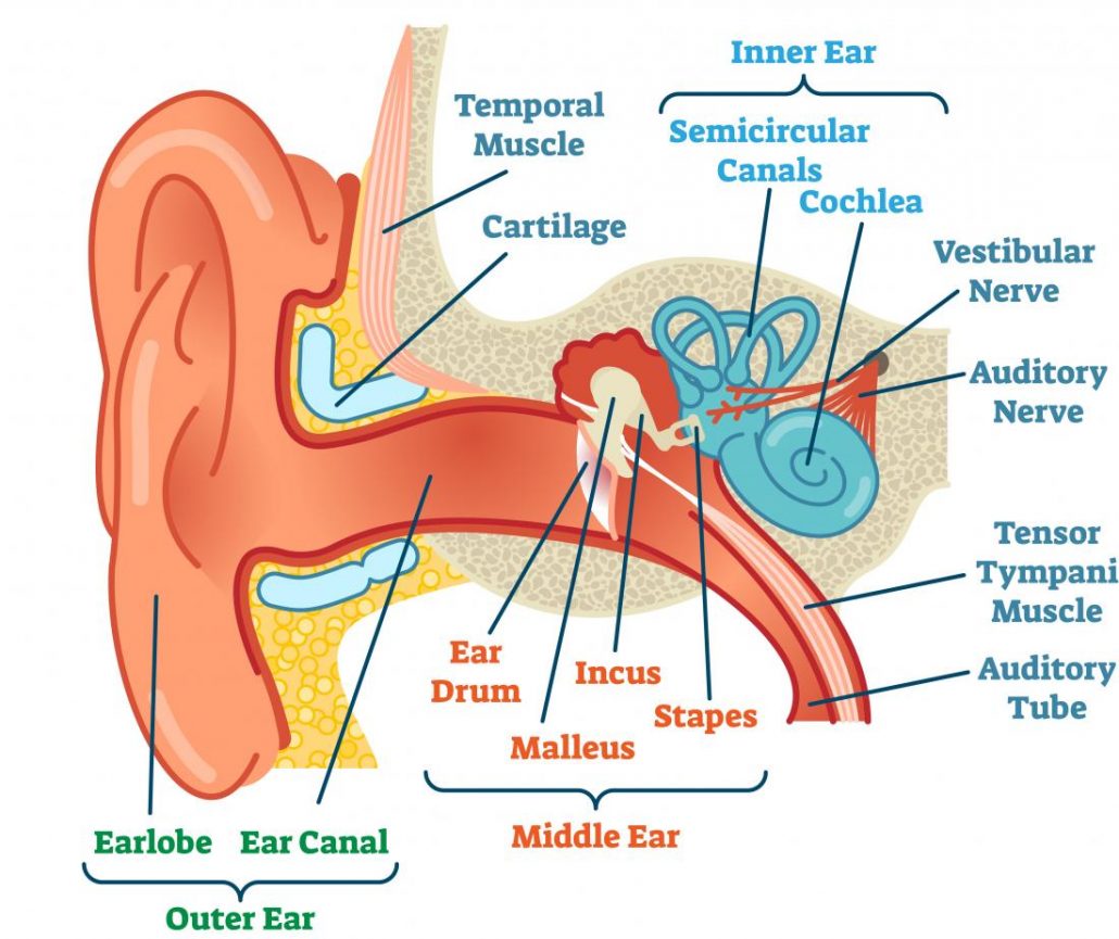
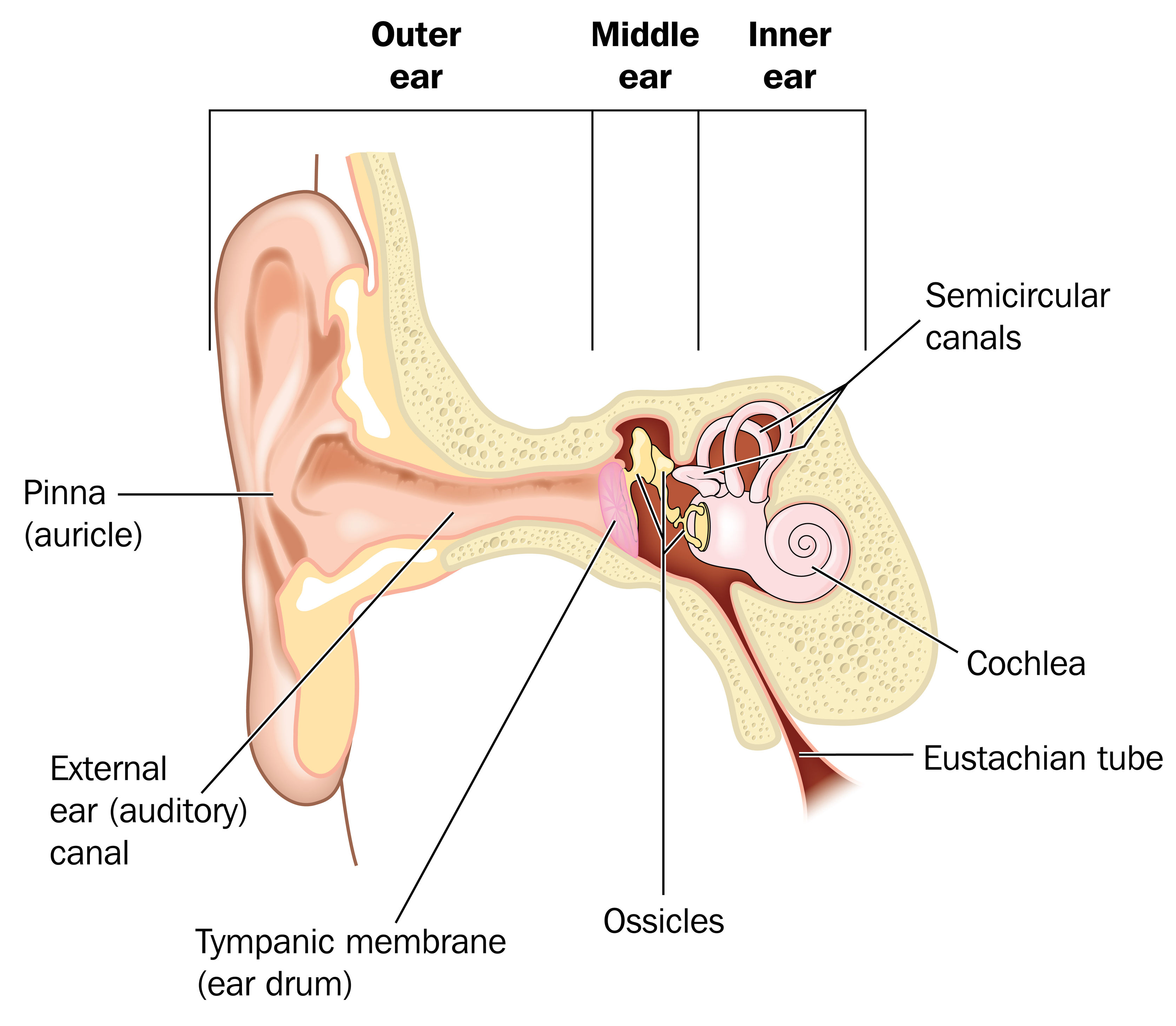
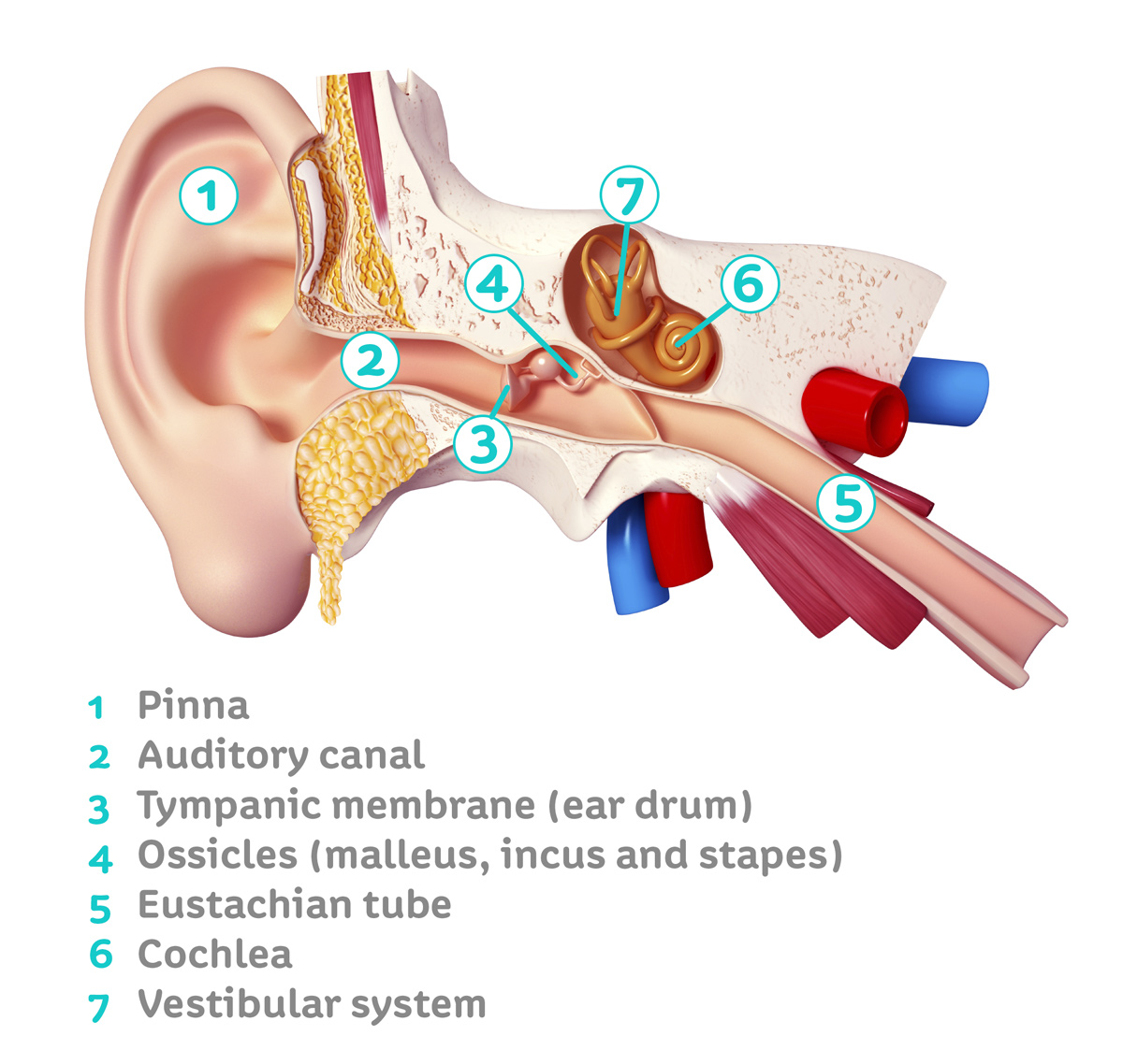
Your outer ear and middle ear are separated by your eardrum, and your inner ear houses the cochlea, vestibular nerve and semicircular canals (fluid-filled spaces involved in balance and hearing). What is the ear? Your ears are organs that detect and analyze sound. Located on each side of your head, they help with hearing and balance. Advertisement
Ear Health Irrigation and Microsuction
How Do We Hear? Hearing depends on a series of complex steps that change sound waves in the air into electrical signals. Our auditory nerve then carries these signals to the brain. Also available: Journey of Sound to the Brain, an animated video. Source: NIH/NIDCD

Functions Of An Ear Inner Ear Parts And Functions Structure And Function Of Inner Ear Human
Ear Anatomy | Inside the ear | 3D Human Ear animation video | Biology | Elearnin Ear is that part of the human body that detects sound from the environment a.

EarQ Anatomy of the Ear Chart Human ear, Inner ear diagram, Ear anatomy
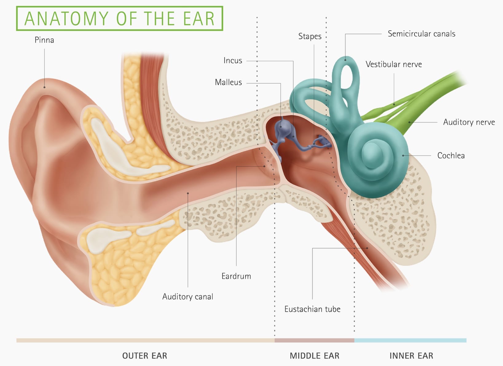
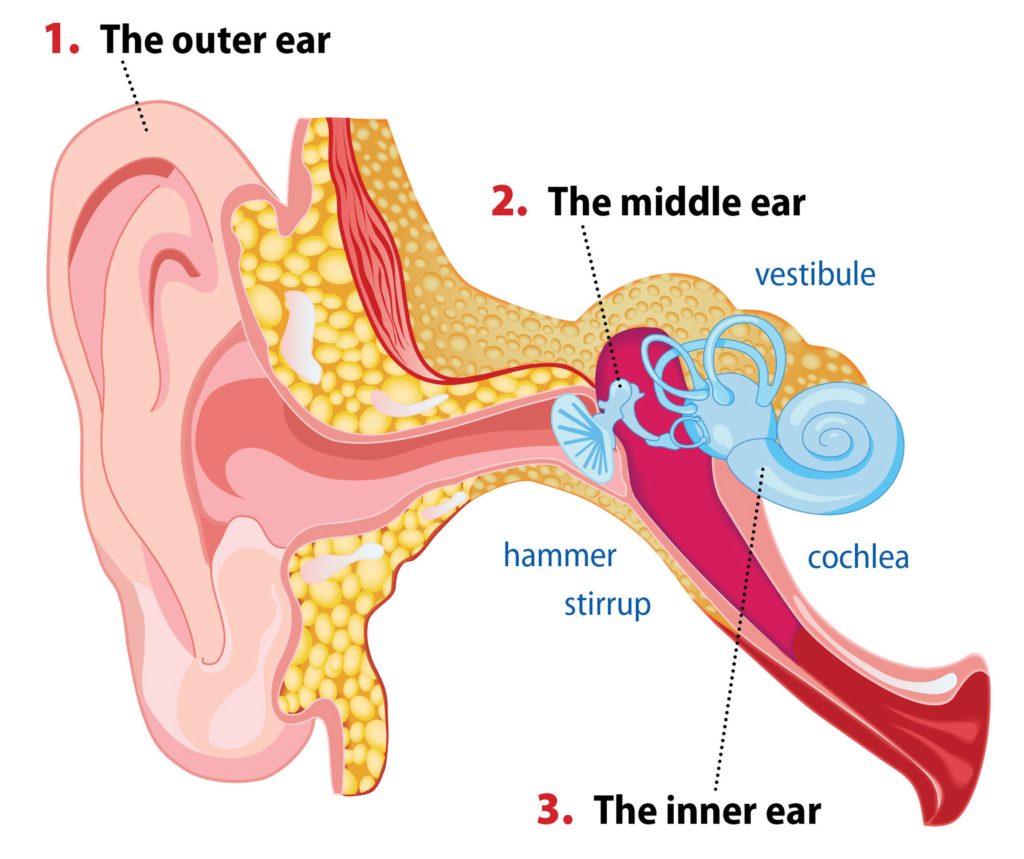
The ear is anatomically divided into three portions: External ear Middle ear Internal ear This mixture of bones, nerves, vessels, membranes, and muscles that make up the ear will be described in this article. Contents External ear Auricle External acoustic meatus Tympanic membrane Muscles of the external ear Vasculature of the external ear
Inner Ear Problems Causes & Treatment of inner ear Dizziness & Vertigo
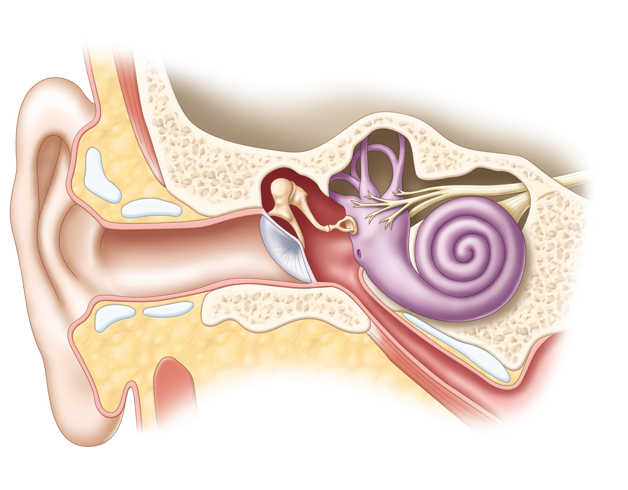
The middle ear (also known as the tympanum or tympanic cavity) is a complicated network of tunnels, chambers, openings, and canals mostly inside openings within the temporal bone on each side of the skull. The 2 largest chambers are called the middle ear space and mastoid.
How The Ear Works Step by Step Brief Explanation
human ear, organ of hearing and equilibrium that detects and analyzes sound by transduction (or the conversion of sound waves into electrochemical impulses) and maintains the sense of balance (equilibrium). Understand the science of hearing and how humans and other mammals perceive sound How humans and other mammals perceive sound.
Inner Ear anatomy Christine Kenney
The inner ear is embedded within the petrous part of the temporal bone, anterolateral to the posterior cranial fossa, with the medial wall of the middle ear, the promontory, serving as its lateral wall.The internal ear is comprised of a bony and a membranous component. The bony part, known as the bony (osseous) labyrinth, encases the membranous part, also known as the membranous labyrinth.
What is conductive hearing loss? Blog of Kiversal
The ear can be divided into three parts; external, middle and inner.This article will focus on the anatomy of the external ear - its structure, neurovascular supply and clinical correlations. The external ear can be divided functionally and structurally into two parts; the auricle (or pinna), and the external acoustic meatus - which ends at the tympanic membrane.
Ear infections explained Dr Mark McGrath
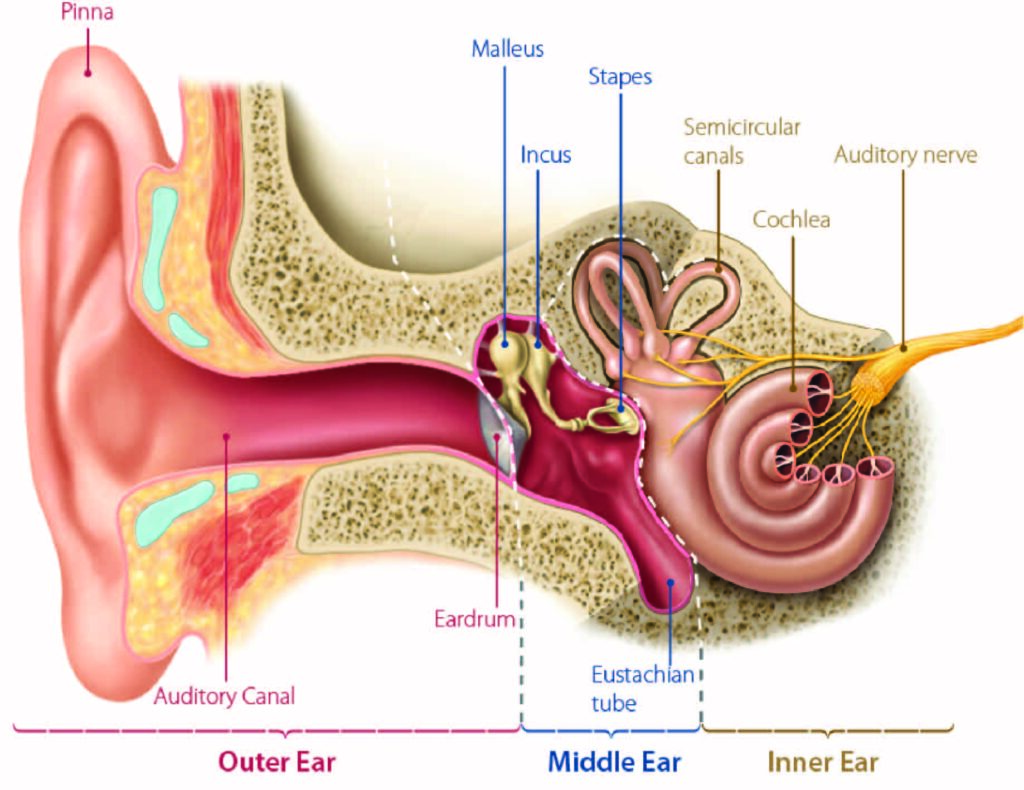
What is the inner ear? What we think of as the "ear" is actually a three-part structure. The outer ear is the part you see and your ear canal. The middle ear is a box-shaped area behind the tympanic membrane (eardrum) that includes the three smallest bones in your body.
Itchy Ears Inside Ear Canal Meaning, Causes, Allergies, Treatment & Superstitions American
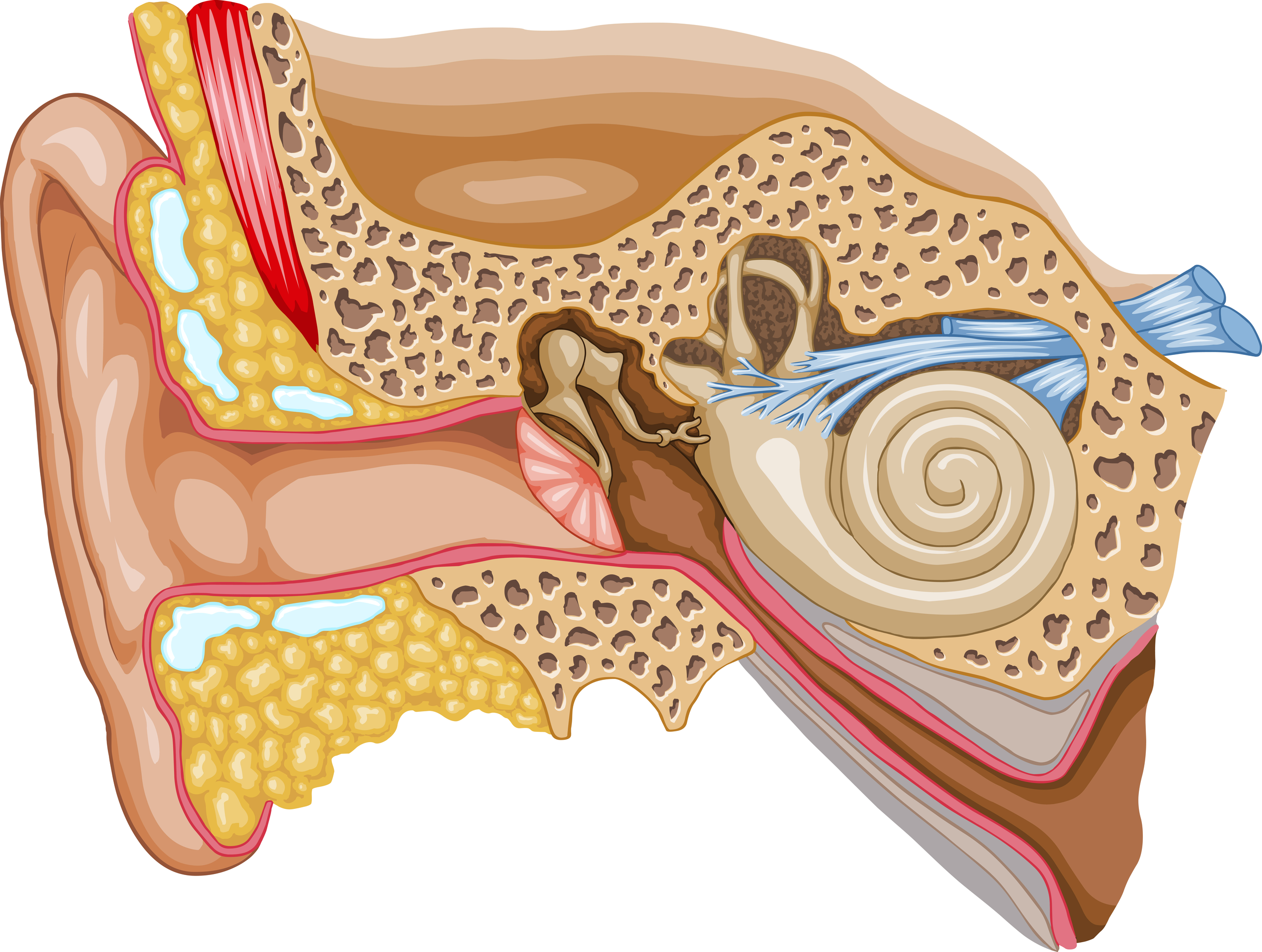
The fluid inside the cochlea, which is a spiral-shaped, fluid-filled structure in the inner ear, is called perilymph. Perilymph is one of the two types of fluid found in the cochlea, the other being endolymph.. So our ear can be divided into three parts: the outer ear, the middle ear, and the inner ear. The outer ear starts with the pinna.
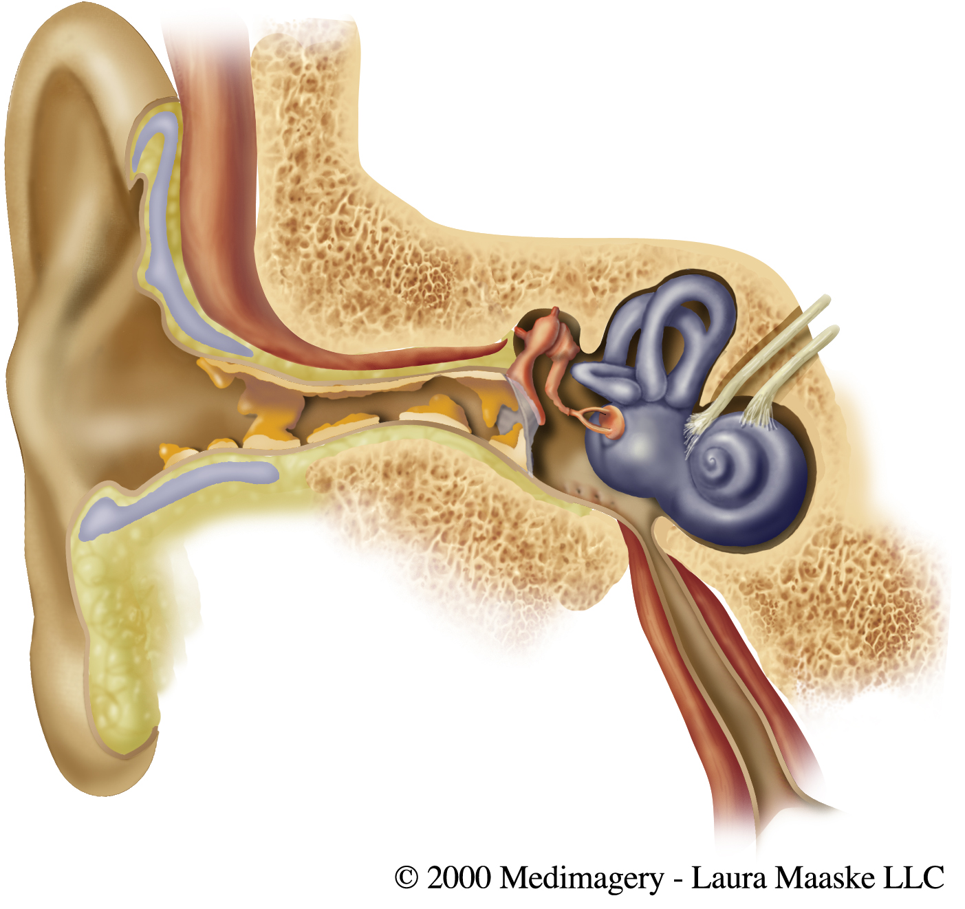
Medical Illustrations Laura Maaske Medical Illustrator & Biological Animator
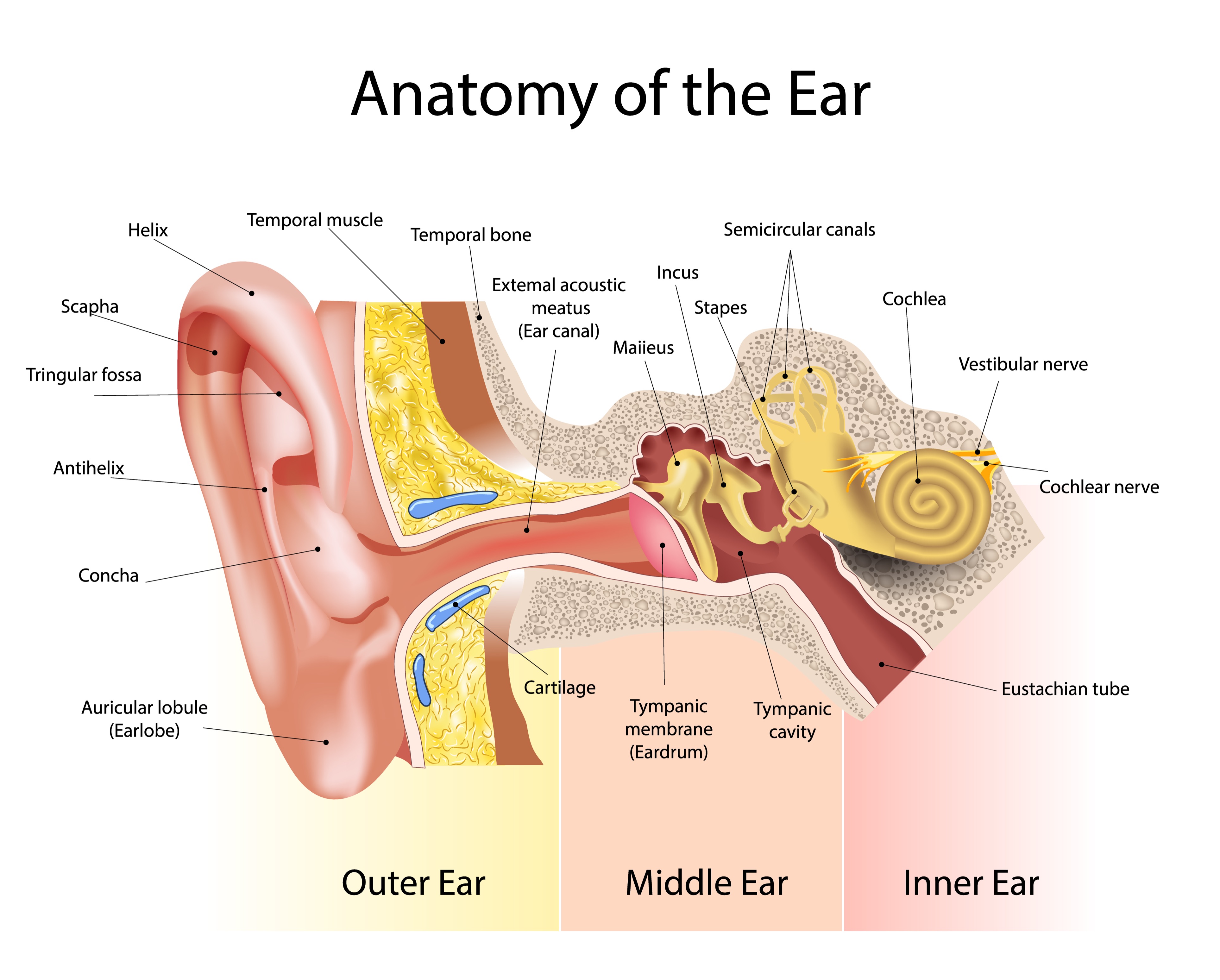
The Structure of Human Ear Helix: It is the prominent outer rim of the external ear. Antihelix: It is the cartilage curve that is situated parallel to the helix. Crus of the Helix: It is the landmark of the outer ear, situated right above the pointy protrusion known as the tragus.
Medical problems of the eyes, ears, nose, and throat symptoms, sinus infection
Inner Ear - Diagram and Description. The human ear comprises three parts, namely the external, middle and inner ear. The inner ear or labyrinth is the innermost part that consists of the bony and membranous labyrinth. The vestibular apparatus is a part of the inner ear that plays a vital role in maintaining equilibrium and posture.
The Ear — Summerlin Audiology
What Is the Anatomy of an Ear? The ear is an unusually complex organ in human anatomy. Don't worry, though—each part has a purpose that is easy to understand. In this section, we describe the anatomy of the ear in simple terms. External Ear Anatomy (Auricle or Pinna)
How You Hear Northland Audiology
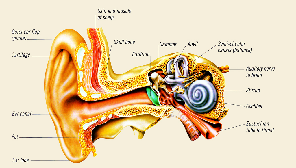
Human ear - Anatomy, Hearing, Balance: The most-striking differences between the human ear and the ears of other mammals are in the structure of the outermost part, the auricle. In humans the auricle is an almost rudimentary, usually immobile shell that lies close to the side of the head. It consists of a thin plate of yellow elastic cartilage covered by closely adherent skin.