Kerley lines wikidoc
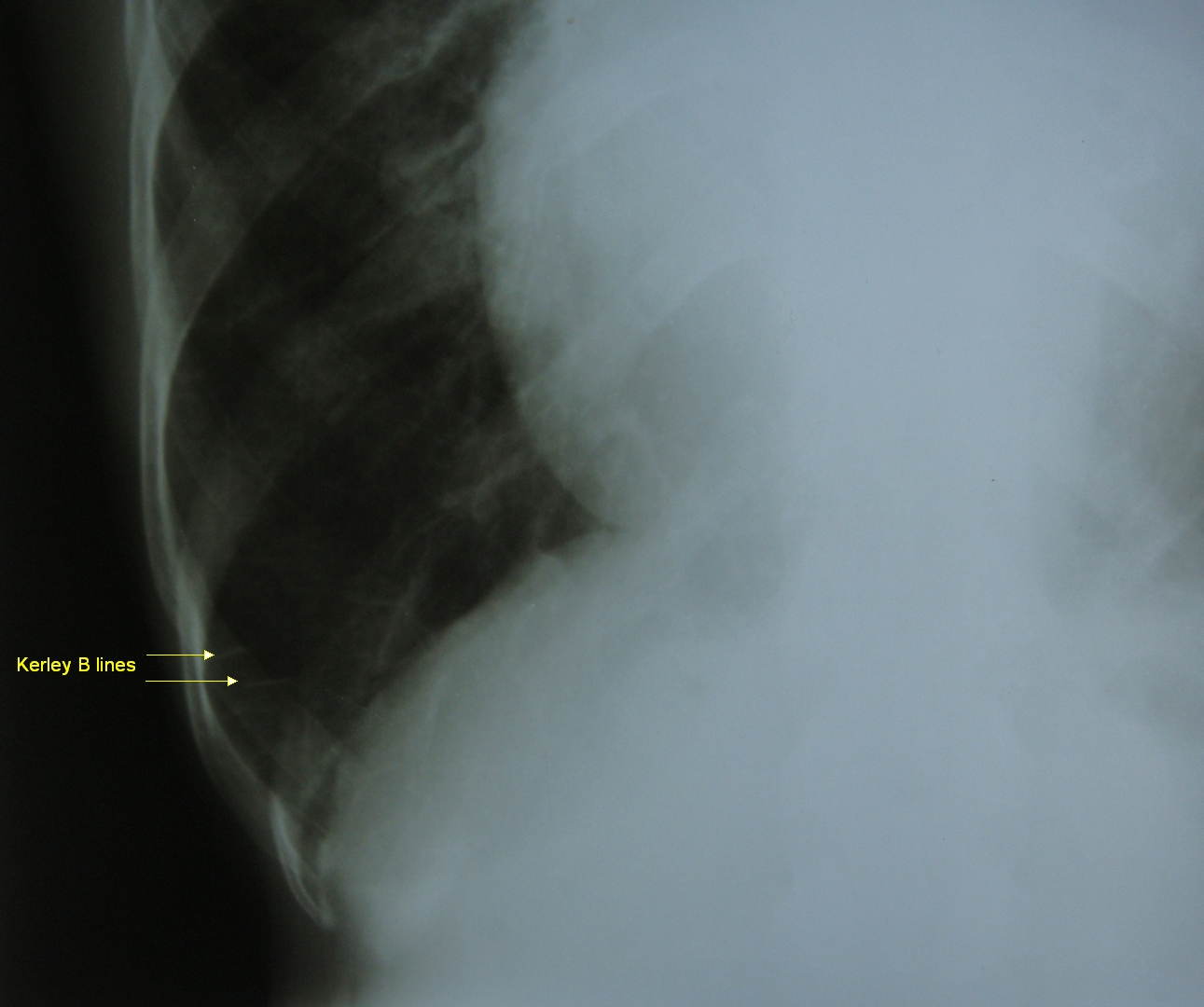
Kerley B lines are linear opacities seen on the chest radiograph. They are 1-2 cm long horizontal lines which meet the pleura at right angles. They are typically seen as a ladder up the side of the lungs beginning at the costophrenic angle. Kerley B lines represent interlobular lymphatics which have been distended by fluid or tissue.
Case 11
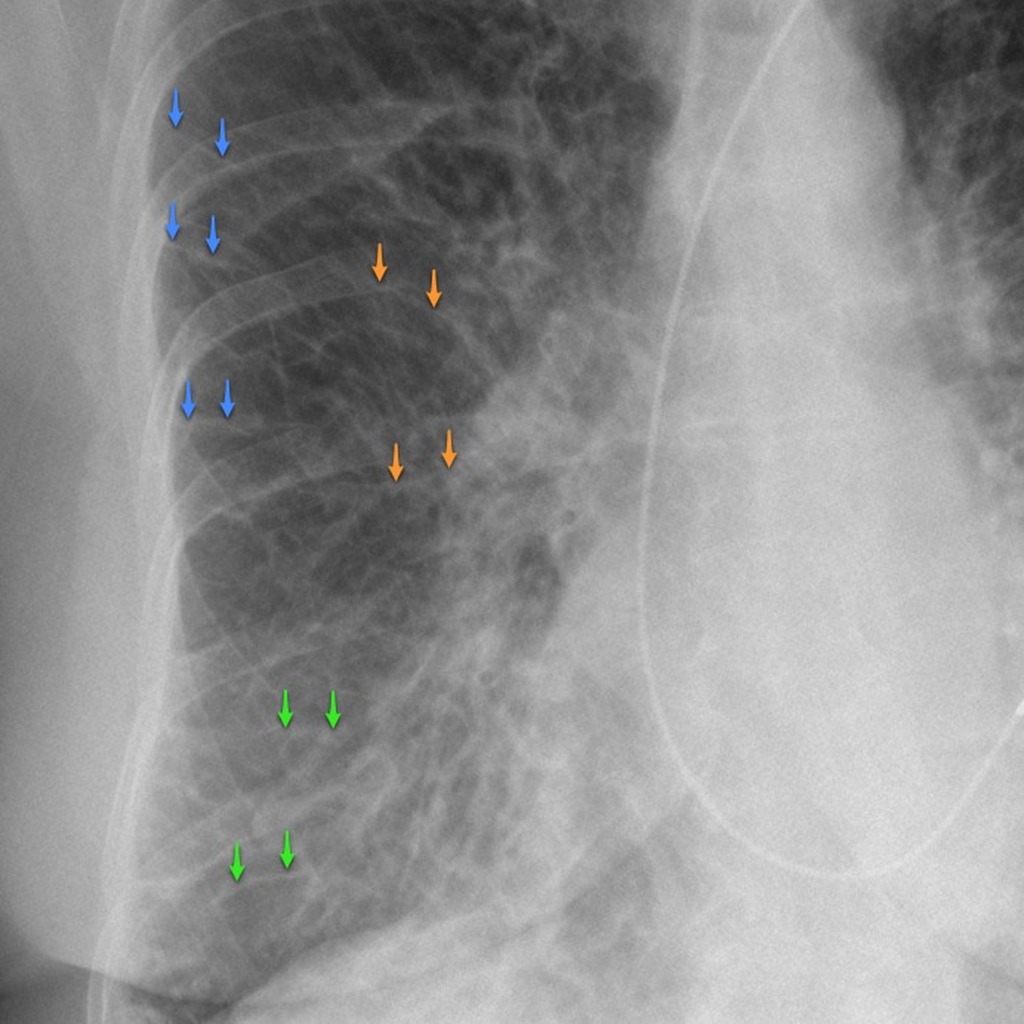
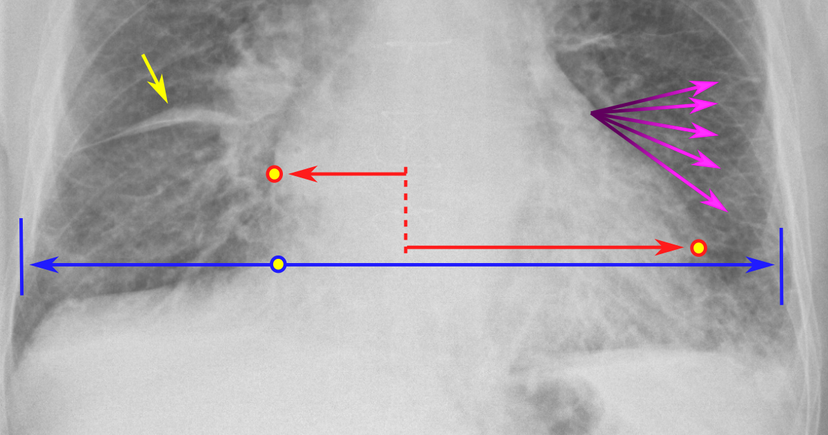
Interlobular septal thickening (Kerley B lines) in the lung apex is a specific sign for pulmonary congestion, although not exclusive (since in ILD there may be apical reticulation). In combination with peribronchial cuffing and increased cardiothoracic ratio, it allows differentiation between cardiac/renal insufficiency and pulmonary ILD.

MD's notebook August 2012
Case Discussion. The septal lines and ground-glass opacity are most suggestive of pulmonary edema. Be sure to look at the PA chest X-ray and the coronal CT images to better appreciate the appearance of Kerley B lines. Normal heart size is compatible with pulmonary edema. Diastolic dysfunction and non-cardiogenic pulmonary edema are examples of.
Acute pulmonary edema finding blue arrow indicates Kerley Blines and... Download Scientific
Kerley's A lines, which radiate 2 to 4 cm from the hilum toward the pulmonary periphery and particularly toward the upper lobes ( Fig. 84-3 ), reflect thickening of the axial interstitial compartment and can be a feature of left ventricular failure or allergic reactions. Kerley's B lines, which reflect thickening of the subpleural interstitial.

Kerley Lines Heart
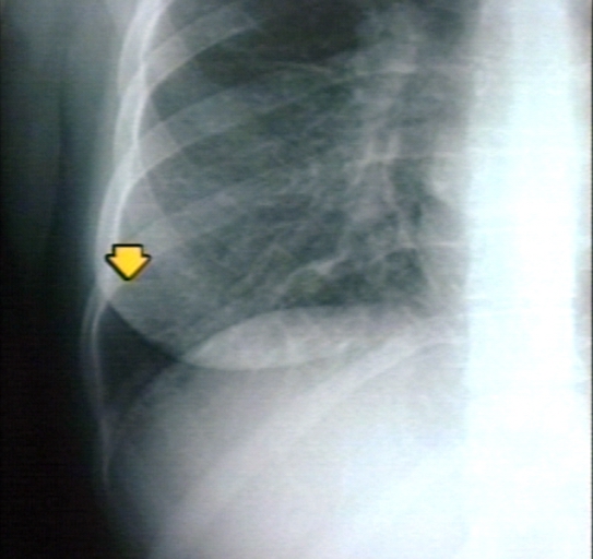
Kerley B lines are short parallel lines at the lung periphery. These lines represent distended interlobular septa, which are usually less than 1 cm in length and parallel to one another at right angles to the pleura. They are located peripherally in contact with the pleura, but are generally absent along fissural surfaces. They may be seen in.
Chest xray showing interstitial lung edema with Kerley B Lines... Download Scientific Diagram
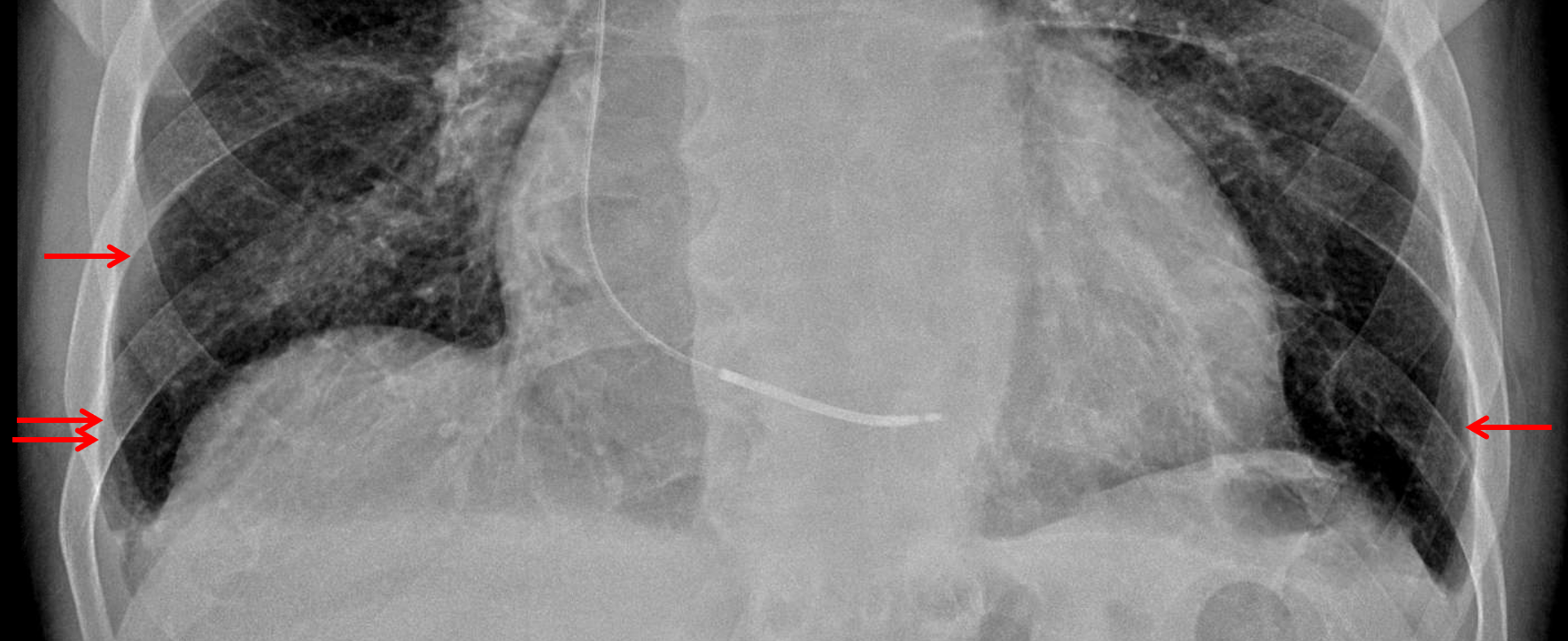
Kerley B-lines: These occur in the area of the pulmonary periphery of the middle lobe, the lingula and the lower lobe. In most cases they are found in the costophrenic angle. They are thin and horizontal lines of about 1 - 2 cm length. They appear as soon as the mean pressure in the left atrium exceeds 20 mmHg at rest [Kasper 2015].
PPT Back to Basics Radiology 2010 PowerPoint Presentation, free download ID4201054
Kerley B lines are small, horizontal, peripheral straight lines demonstrated at the lung bases that represent thickened interlobular septa on CXR. They represent edema of the interlobular septa and though not specific, they frequently imply left ventricular failure. Kerley C lines are reticular opacities at the lung base, representing Kerley.
Kerley B Lines Cxr Septal lines in lung Radiology Reference Article They are thin
What are Kerley B Lines? We created this video to cover the medical definition and provide a brief overview of this topic.💥Kerley B lines [Full Guide].
cxr stase Kerley B lines [This website has no commercial interest Only personal reflections]
Plain radiograph. There are bilateral basal interstitial lines that extend to the pleural surface - these are septal (Kerley B) lines. There is slight asymmetry of the breast shadows and metallic clips in the right axilla. Features are consistent with previous breast carcinoma and lymphangitis carcinomatosis. I would compare this with previous.

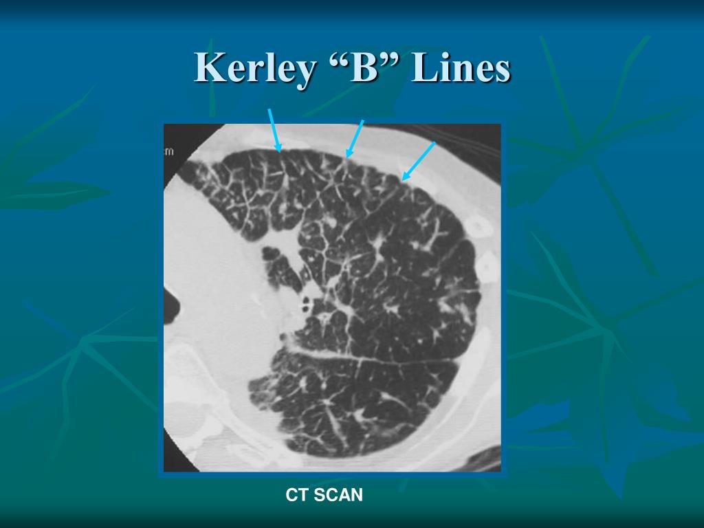
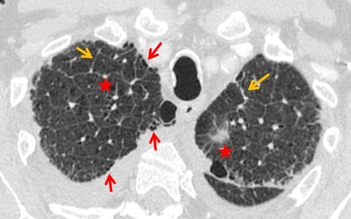
Kerley B lines on CT Image
Kerley B Lines Kerley B lines (arrows) are horizontal lines in the lung periphery that extend to the pleural surface. They denote thickened, edematous interlobular septa often due to pulmonary edema.

PPT Diagnostic Radiology Congestive Heart Failure PowerPoint Presentation ID6637651
Citation, DOI, disclosures and article data. Septal lines, or Kerley lines, are seen when the interlobular septa in the pulmonary interstitium become prominent. It may be because of lymphatic engorgement or edema of the connective tissues of the interlobular septa. They usually occur when pulmonary capillary wedge pressure reaches 20-25 mmHg.
Kerley B lines Chest Xray « PG Blazer
Heart Failure Kerley B lines. In these images. a nd c are normal and b and d represent thickened interlobular septa in a patient with congestive heart failure. These are the well known Kerley lines, often spoken about but rarely seen. They are identified as thin horizontal lines usually seen in the costophrenic angles, not being longer than.

Kerley B (septal) lines Image
Kerley B lines (thickened interlobular septa) are much spoken about as a medical student, but less commonly observed than one might expect given the volume of cardiac failure patients. These thin lines of 1-2 cm are virtually always at the lungs bases and at the lung periphery lying perpendicular to the pleural surface to which they contact.

Image Kerley B Lines Merck Manuals Professional Edition
Hey guys! Dr Sharma DO here!Quick lesson on Kerley B Lines, and just overall how to interpret a chest xray that is suggestive of heart failure. Like and Subs.
Kerley B Lines Cxr Septal lines in lung Radiology Reference Article They are thin
Kerley B lines in a patient with congestive heart failure. Kerley B lines. These are short parallel lines at the lung periphery. These lines represent interlobular septa, which are usually less than 1 cm in length and parallel to one another at right angles to the pleura. They are located peripherally in contact with the pleura, but are.
Kerley B lines in the lung apex a distinct CT sign for pulmonary congestion
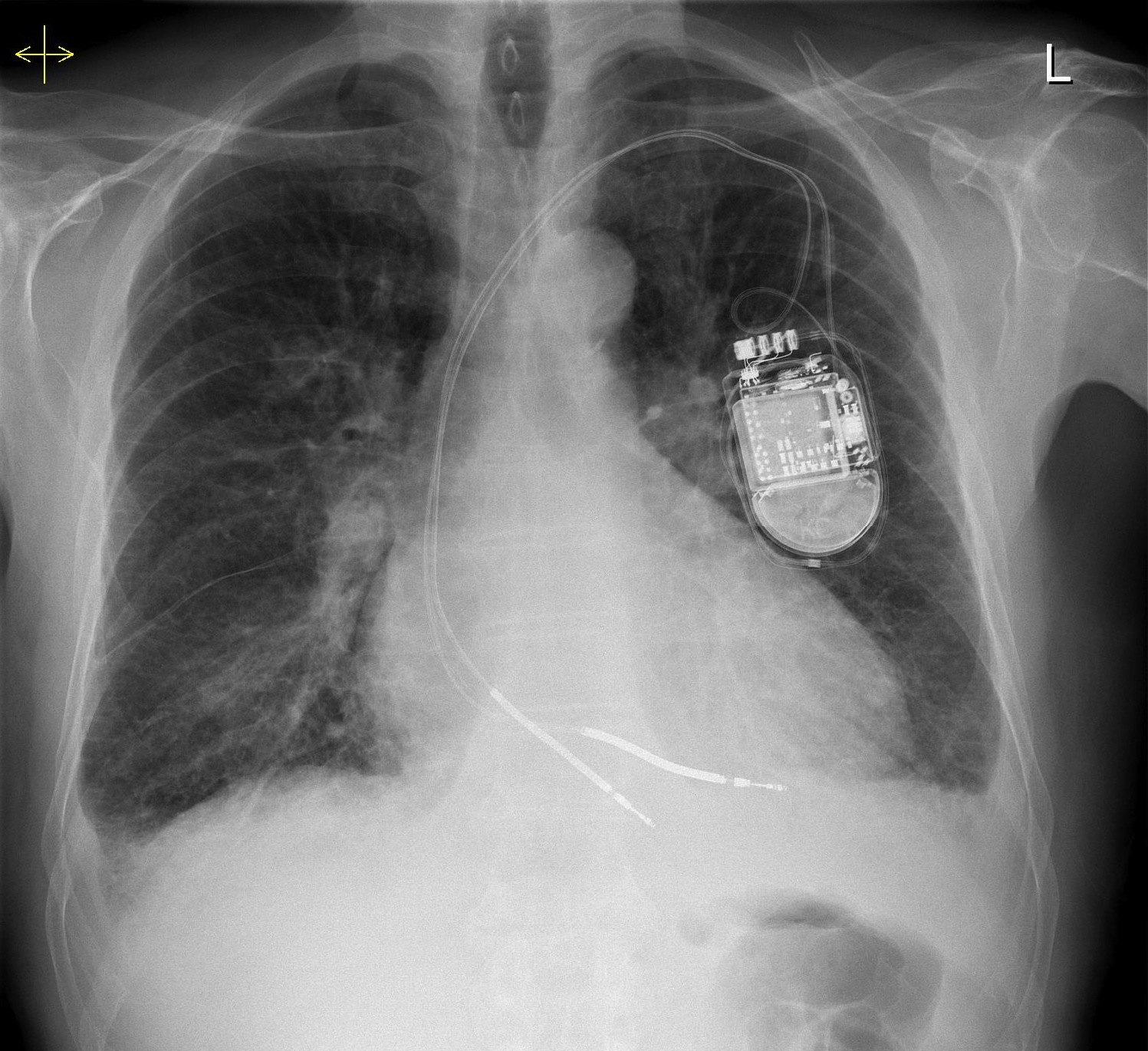
Chest x-ray 10 days earlier. x-ray. Single lead permanent pacemaker (PPM) in situ. Heart is enlarged, and there is some prominence of the pulmonary vasculature, without evidence of interstitial or alveolar edema.