
Tibia Pelvic limb bone anatomy Horse Veterinary YouTube
#6. Tibia is the larger and longer bone of a horse. #7. There is a grooved anterior tuberosity in the horse tibia bone #8. There is six tarsal bone found in horse - tibial tarsal, fibular tarsal, central tarsal, first and second fused tarsal, third and fourth tarsal bones. #9.

Radius and Tibia of a Horse, Vintage Engraving Stock Vector
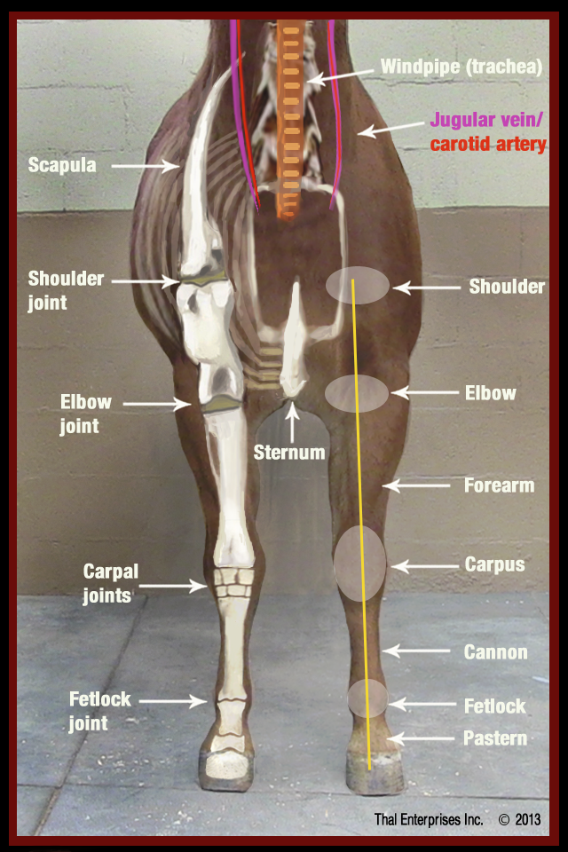
• Metacarpals - there are three metacarpal bones in the horse, which may be numbered 2-4. Of these the third or large metacarpal,. • Tibia and fibula - the tibia runs down diagonally from the femur to the hock and is the larger of the two bones allowing for the attachment of the major muscles responsible for the movement of the.
Horse tibiaCut marks and fragmentation in layer 5. Download
In most light horse breeds a cannon bone circumference that is greater than 8 inches is desirable. This means the horse has a sturdy bone mass to carry a load and withstand work. These bones are somewhat equivalent to the metacarpal bones in a human's palm.. The underlying bones are the tibia and the smaller fibula which are equivalent to.
Fracture of Radius or Tibia Horse Side Vet Guide
In the horse, these development periods are completed very early in life, generally by 2 years of age. Using a variety of measures to define the completion of growth and bone development, the horse enters skeletal maturity by the time it is 2 years old. There is little variation in the age of maturity across different horse breeds.
Fracture of Radius or Tibia Horse Side Vet Guide
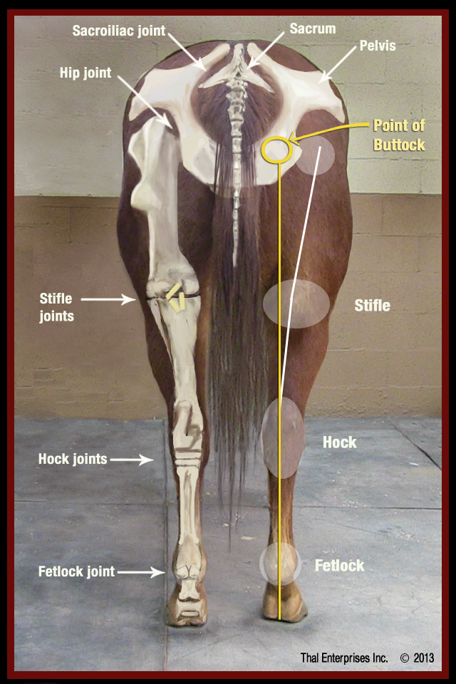
When working with horses, it is important to be able to accurately assess, diagnose and manage an equine patient. To do this, a good understanding of equine anatomy is essential. Anatomy [edit | edit source] Pelvic hind limb bears 40-45% of the weight and provides the majority of propulsion for locomotion. Bones [edit | edit source]
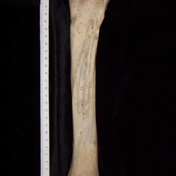
Horse (Equus caballus) left tibia, posterior view BoneID
Horse - Digital bones of the hand: Proximal phalanx [Long pastern bone], Middle phalanx [Short pastern bone], Distal phalanx [Unguicular bone, Ungual bone], Medial ungular cartilage, Distal sesamoid bone Horse - Coxal bone: Acetabulum, Ilium, Ischium, Pubis Veterinary anatomy - Thigh bone [Femur] (Horse) Horse: Tibia-Fibula Horse - Skeleton of.

. The surgical anatomy of the horse Horses. Plate XIII.—The Right
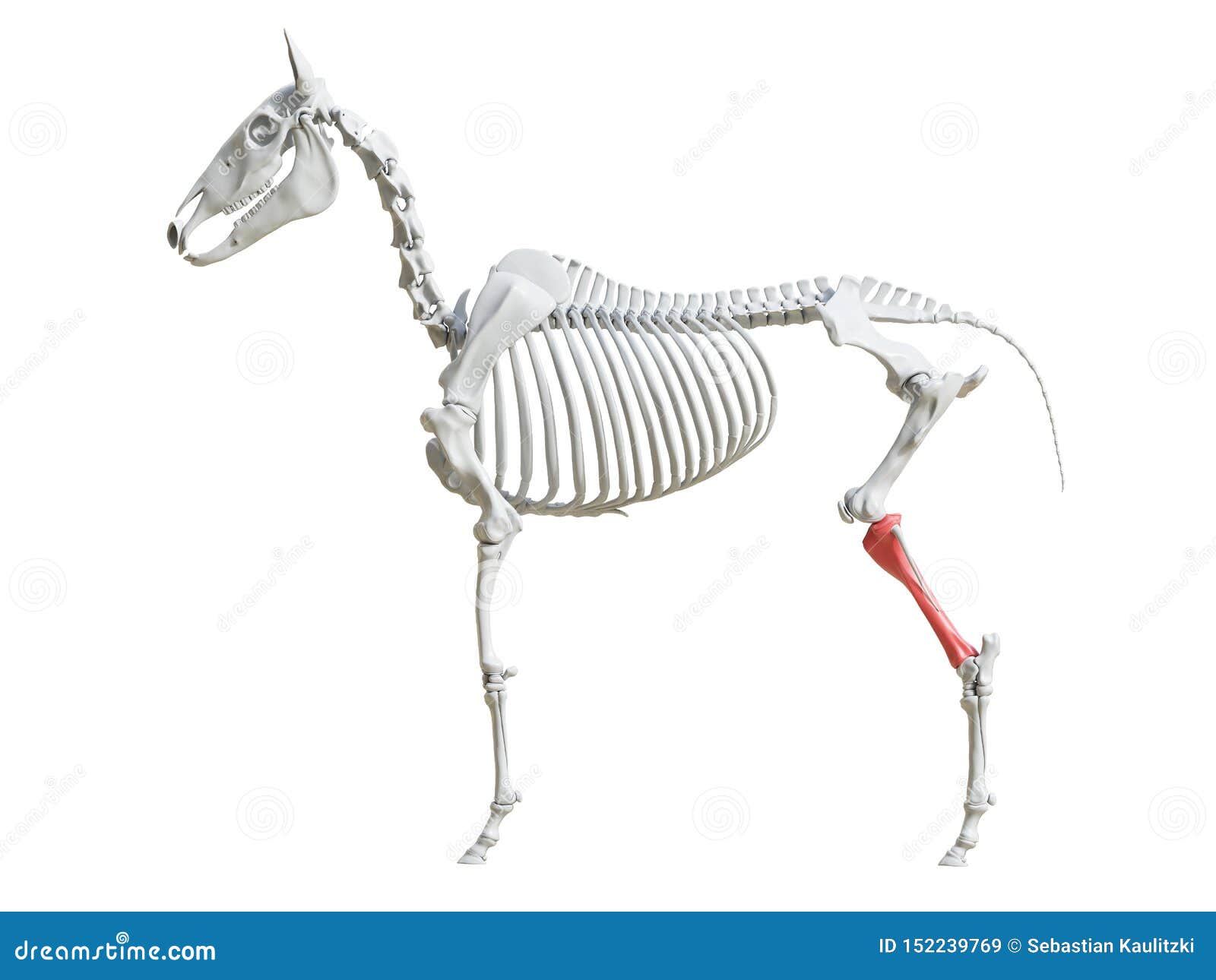
The femur, which is a large bone, connects with the pelvis at the hip joint and with the hind leg at the stifle joint. The tibia forms the upper part of the hind limb from the stifle to the hock. The fibula is a smaller bone that extends half the length of the tibia and sits parallel to it.
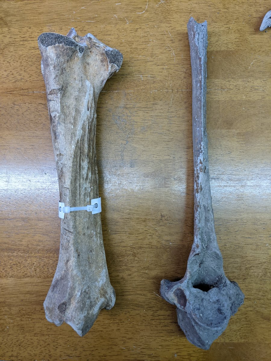
Horse Tibia and Bovid Vertebrae Members Gallery The Fossil Forum
Bucked Shins/Dorsal Cortical Fractures of the Third Metatarsal Bone in Horses.. Osseous cyst-like lesions may also occur in the proximal tibia. The pathogenesis of these cysts is poorly understood, but they may develop after trauma to the articular surface or as a result of osteochondrosis. Lesions often present in young horses but can be.

Horse (Equus caballus) left tibia, proximal articular surface BoneID
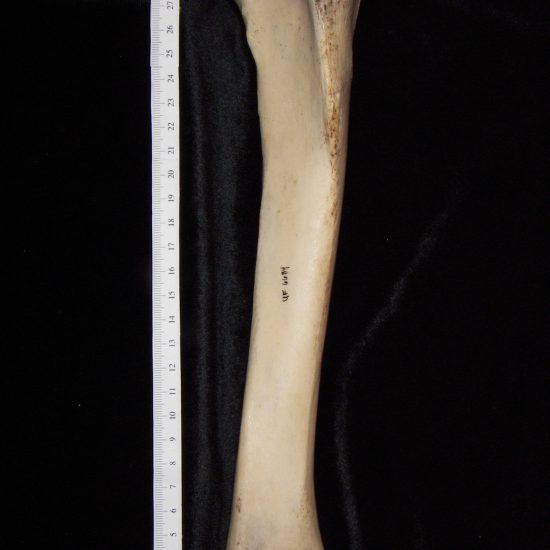
Tibia. The horse's tibia is a long bone and is present between the stifle joint and the hock joint. The upper end of the tibia provides the place for the junction of the muscles in the hock and the lower limb.. The horse's fibula bone is so small that it is almost vestigial. Tarsal bones. The tarsus also known as hock is made up of six.

Pin by Mladen Djokovic on Horse (Bones) Horse bones, Horses, Bones
The bones that mostly determine the horse's height (cannon, tibia, femur, scapula) will generally stop growing around 4 years of age. But some bones that still add to the horse's height, such as the spine tips at the withers , will continue to grow until 5 or 6 years of age.

Horse Leg Bones Diagram / Hoofnotes Infographic Equine Anatomy Part 4
Learn about the veterinary topic of Fracture of the Lateral Malleolus of the Tibia in Horses. Find specific details on this topic and related topics from the MSD Vet Manual.. Focal Bone Reaction and Avulsion Fractures of the Third Metatarsal Bone in Horses. Fractures of the Second and Fourth Metatarsal Bones in Horses. Enostosis-like Lesions.
Horse (Equus caballus) left tibia, distal articular surface, view 2
They are bones of similar design, so they are discussed together here. A hairline non-displaced fracture of the radius or tibia may cause severe lameness. These fractures usually result from a kick, but can happen with certain types of overload as well. Horses with hairline fractures must be confined for 8 weeks until the fracture is healed.
The Equine Skeleton Tibia Stock Illustration Illustration of tibia
Tibia: leg bone. Calcaneus: bone that forms the hock tip. Tarsus: bone forming the joint between the tibia and the metatarsus. Metatarsus: hock bone.. depends on the horse's size, breed, gender, and the quality of care provided by its owner. Also, if the horse is larger, its bones are larger; therefore, not only do the bones take longer to.
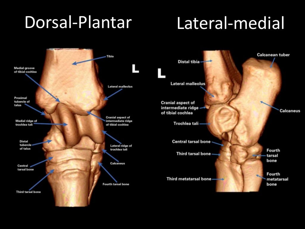
Tarsal Anatomy of the Horse
The hock, also known as the tarsus, joins the upper leg and lower leg together - it attaches the tibia to the cannon bone (a horse's shin). The hock is a complex area - it consists of 4 different joints and gives your horse the power to spring over fences. Horse Leg Anatomy - Upper Forelegs

Osteochondrosis, OC, Osteochondritis Dissecans, OCD Horse Side Vet Guide
Its medial surface presents a narrow area along the proximal border for articulation with the lateral condyle of the tibia bone. The distal extremity of the fibula is fused with the tibia bone. Now, I would like to summarize the special features of tibia and fibula bones of horse - The tibia of a horse is a larger and longer bone in the skeleton.

Horse TibiaFibula Horse anatomy, Anatomy, Horses
A horse has 205 bones, which is why equines have a majestic build. It took millions of years of evolution for horses to have elongated bones.. where it contacts the patella and tibia. Tibia - This bone is from the stifle to the hock. It has a proximal end that provides a bond for some ligaments. They hold collateral ligaments, cruciate.