
Bochdalek hernia On conventional radiographs, the hernia may appear as a
Bochdalek hernia is a developmental defect in the posterolateral diaphragm, allowing herniation of abdominal contents into the thorax, causing mechanical compression of the developing lung parenchyma and sometimes causing lung hypoplasia. As such, symptoms typically manifest in the pediatric age group and tend to be respiratory.
Bochdalek (Pleuroperitoneal) Hernia radRounds Radiology Network
The Bochdalek hernia was confirmed on computed tomography (CT) imaging and the patient underwent surgical repair with Gore-Tex mesh. Conclusion The report shows a rare case of the Bochdalek hernia in a young adult, successfully managed with a laparotomy.

Bochdalek Hernia images, diagnosis, treatment options, answer review Thoracic Imaging
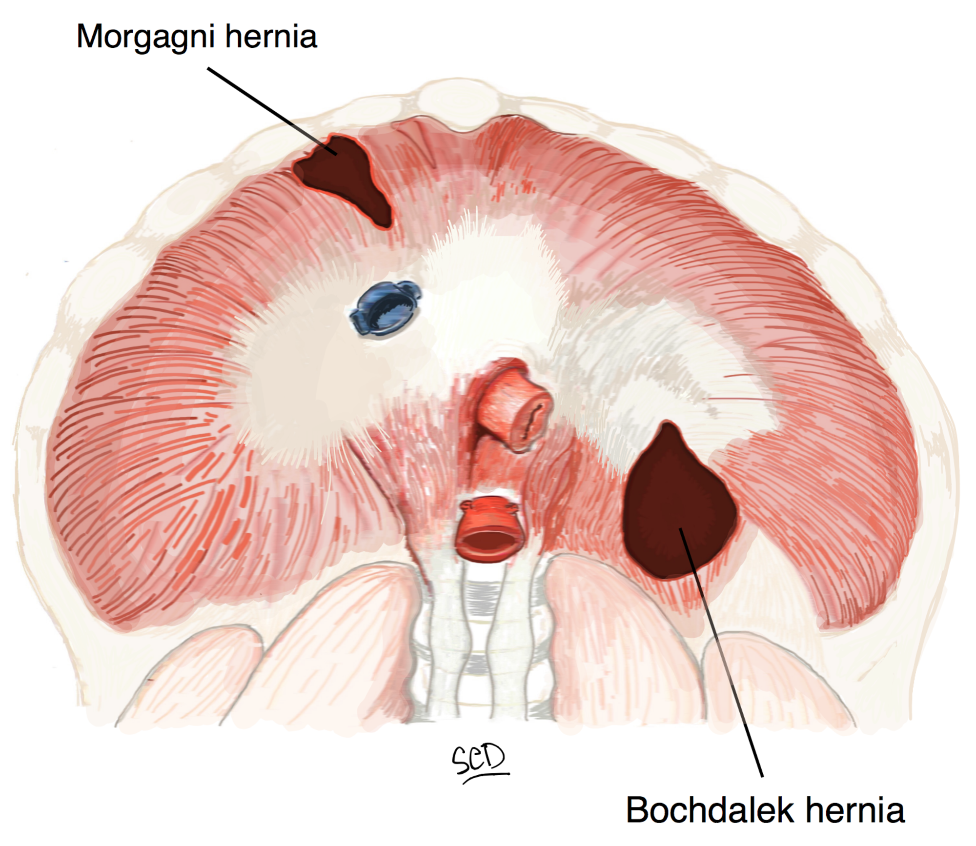
Bochdalek hernia is one of two forms of a congenital diaphragmatic hernia, the other form being Morgagni hernia. A Bochdalek hernia is a congenital abnormality in which an opening exists in the infant's diaphragm, allowing normally intra-abdominal organs (particularly the stomach and intestines) to enter into the thoracic cavity.
Learning Radiology Congenital Diaphragmatic Hernia, Bochdalek ,
Bochdalek hernia occurs through defects in the pars lumbaris and pars costalis and is more common on the left side. These are the most common congenital diaphragmatic hernias, with an estimated incidence of 1 per 2000-5000 live births . In adults, Bochdalek hernias are often underreported and can be identified in 0.17% to 6% of patients.

Bochdalek hernia Radiology Case
Bochdalek hernia (BH) is the most common type of congenital diaphragmatic hernia, accounting for 90% of these hernias [1]. BH is more common on the left side (85%) [2]. Late presenting congenital diaphragmatic hernia poses a considerable diagnostic challenge, since this diagnosis in the adult age is extremely rare; being mostly diagnosed during the neonatal period. [3]

Rightsided Bochdalek’s hernia in an adult The American Journal of Surgery
Bochdalek hernia is a posterolateral diaphragmatic defect resulting from failure of the retroperitoneal canal membrane to fuse with the dorsal esophageal mesentery and the body wall. The location of the foramina of Bochdalek is defined by the location of the diaphragmatic coronary ligaments bilaterally. The herniation is termed "congenital.

Neonatal Bochdalek hernia. a A 1dayold boy with left Bochdalek... Download Scientific Diagram
Journal of Thoracic Imaging, Vol. 24, No. 1 Prevalence of Incidental Bochdalek's Hernia in a Large Adult Population November 23, 2012 | American Journal of Roentgenology, Vol. 177, No. 2

Congenital diaphragmatic herniation (CDH) accounts for a small proportion of all diaphragmatic
Abstract The chest and abdominal computed tomography (CT) scans of 940 patients were reviewed to determine the prevalence of Bochdalek hernias and to evaluate the widely held concept that left-sided hernias occur more than nine times as often as right-sided hernias.
Cureus Diaphragmatic Hernia Repair Using Biosynthetic Tissue Reinforcement Patch A Case
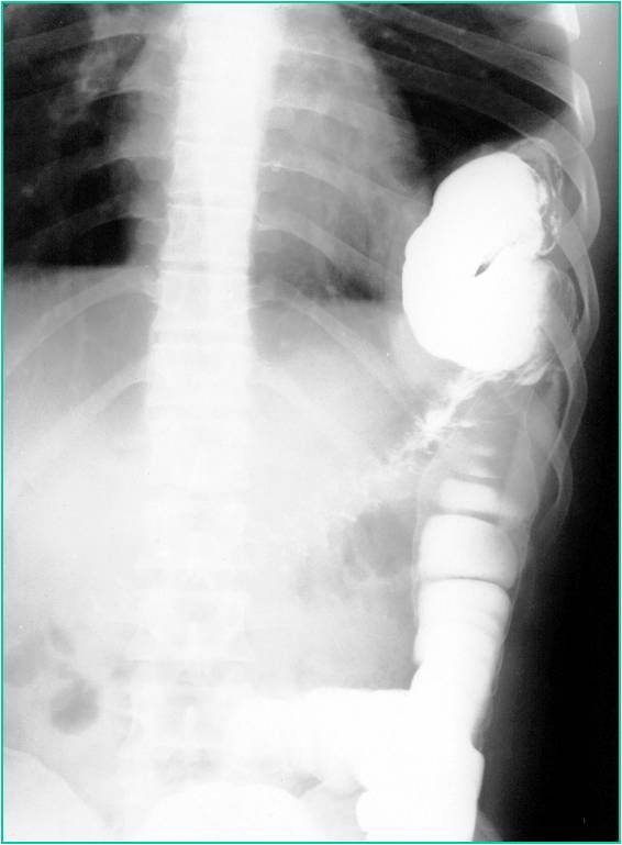
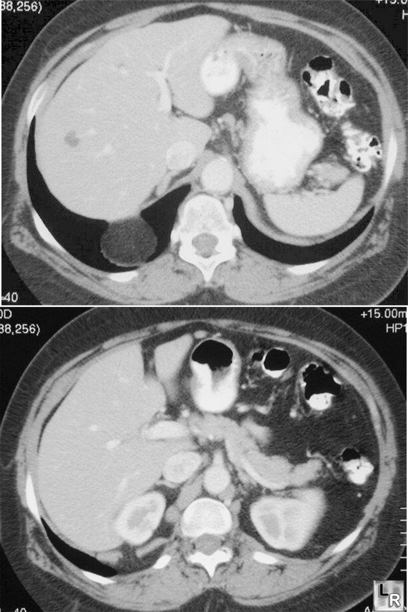
Coronal C+ portal. venous phase. Sagittal C+ portal. venous phase. CT scout image reveals indistinct right diaphragmatic copula with right paracardiac soft tissue shadow. CT images show a defect of the right crus of the diaphragm with herniation of the stomach, the first part of the duodenum and part of the left lobe of the liver into the right.

Image
Bochdalek hernias , also known as pleuroperitoneal hernias, (alternative plural: herniae) are the commonest type of congenital diaphragmatic hernia. They occur posteriorly and are due to a defect in the posterior attachment of the diaphragm when there is a failure of pleuroperitoneal membrane closure in utero.

Bochdalek Hernia images, diagnosis, treatment options, answer review Thoracic Imaging
Bochdalek hernia is a type of congenital diaphragmatic hernia that primarily manifests in children. It is rare in adults and accounts for about 0.17% to 6% of all diaphragmatic hernias [ 1, 2 ]. Bochdalek hernia affects approximately 1 in 2200 to 12,500 live births and was first described by Vincent Alexander Bochdalek in 1848 [ 3 ].
Bochdalek hernia radRounds Radiology Network
Coronal C+ portal venous phase. Sagittal C+ portal venous phase. CT. CT scout. CT scout image reveals indistinct right diaphragmatic copula with right paracardiac soft tissue shadow. CT images show a defect of the right crus of the diaphragm with herniation of the stomach, the first part of the duodenum and part of the left lobe of the liver.

Bochdalek Hernia images, diagnosis, treatment options, answer review Thoracic Imaging
OBJECTIVE. The purpose of this study was to determine the prevalence and characteristics of adult Bochdalek's hernia in a large patient population. MATERIALS AND METHODS. We retrospectively reviewed all abdominal CT scans obtained at our hospital in 1998.

Bochdalek hernia Radiology Case
ct CT thorax coronal view demonstrates a right Bochdalek hernia. There is herniation of peritoneal fat through a congenital diaphragmatic defect, Bochdalek foramen, in the posterolateral right hemidiaphragm. Bochdalek hernia is usually left sided and maybe an incidental finding in as high as 10% of asymptomatic adult 1 .

Symptomatic Bochdalek hernia in an adult patient BMJ Case Reports
Radiological features CXR - a well-defined, dome-shaped soft tissue opacity is seen midway between the spine and the lateral chest wall. This may 'come and go'. There may be loops of bowel or gas-filled stomach within the area. The ipsilateral lung may be smaller with crowding of the bronchovascular markings and occasionally mediastinal shift.

Bochdalek Hernia images, diagnosis, treatment options, answer review Thoracic Imaging
Presentation Shortness of breath. Patient Data Age: 70 years Gender: Female ct Axial lung window Coronal lung window Axial non-contrast There is herniation of some peritoneal fat through a diaphragmatic defect in the posteromedial aspect of the left hemidiaphragm. Case Discussion