
Wave goodbye to pain Foot Anatomy and Biomechanics
The anatomy of a healthy cow's foot Knowledge about cattle foot anatomy may help to understand why lameness occurs. Back to: Lesions of cows' feet It is essential to be able to recognise the different parts of the horn, particularly when foot trimming .
DISTAL LIMB SURGERIES AND HOOF TRIMMING
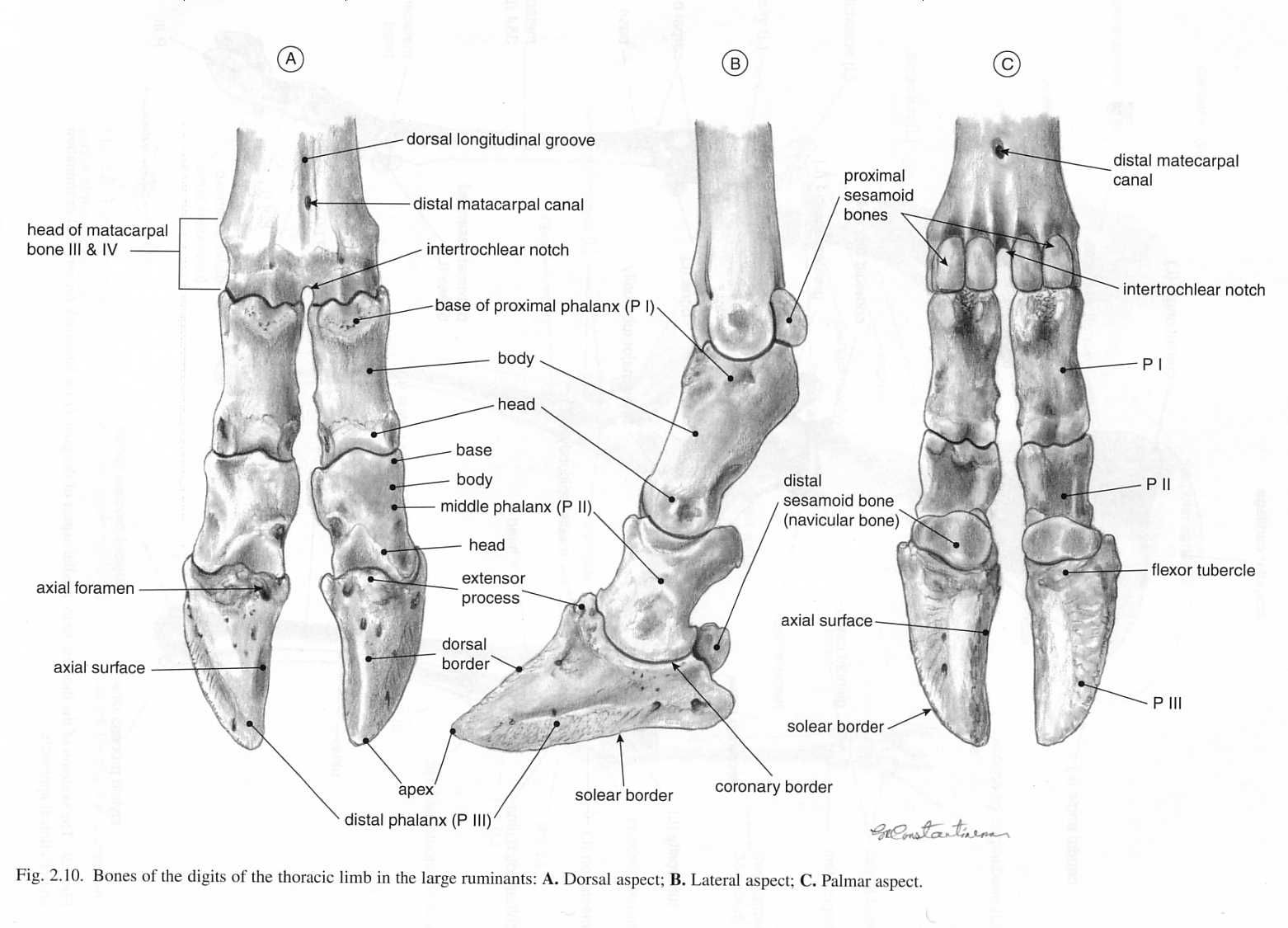
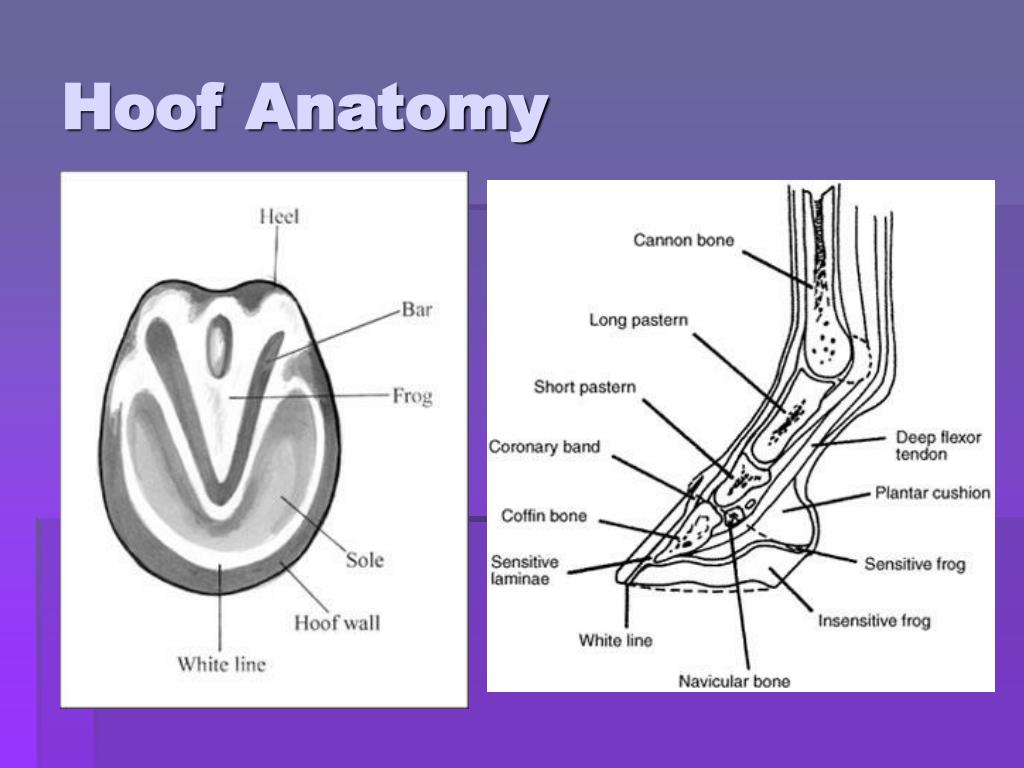
The distal interphalangeal joint (or DIP joint) is the articulation between the middle and distal phalanges. The ventral aspect of the distal phalanx has the flexor tuberosity, where the deep flexor tendon attaches. Continuous pressure by the flexor tuberosity to the corium may result in hemorrhage and ulceration.

The Anatomy of the Hoof Hoofcount
The horse's hoof is a miracle of engineering. It contains a whole host of structures which, when healthy, operate in equilibrium with each other to form a hoof capsule which is able to withstand huge forces, utilising energy to assist with forward movement while providing protection to the sensitive structures beneath.

SheepHoofAnatomy.jpg (800×562) Hooves, Sheep, Goat feet
The distal end of the cannon bone bears a condyle, with an extensive cylindrical articular surface, and a prominent sagittal ridge that articulates with a complementary sagittal groove of the proximal phalanx (P1). (see Fig. 7 image below of fracture related to these sagittal structures)
Farm Health Online Animal Health and Welfare Knowledge Hub Lameness
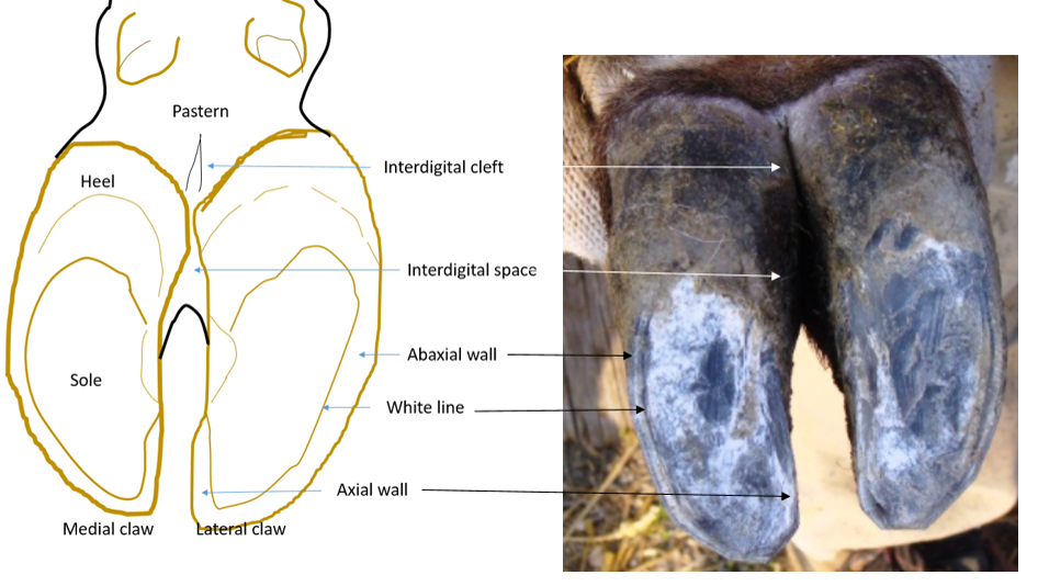
Anatomy of a bovine hoof Cattle are cloven-footed animals and the hoof consists of two digits. The claws are named by their relative location on the foot. There is the outer, or lateral claw, and the inner, or medial claw. The space between the two claws is called the interdigital clef; the area of skin is called the interdigital skin.

Cow Foot with Hoof Natural Specimen Anatomy Model, Articulated
The hoof consists of two parts. The broad, hard, upper portion is called the unguis; it completely surrounds the end of the toe, extending down and forming a rim around the bottom of the hoof. A somewhat softer plate, called the subunguis, covers the bottom of the toe and is extensively developed in hoofed animals to form a tough pad. (In humans the subunguis is only a small ridge under the.

Cow Foot with Hoof Natural Specimen Anatomy Model, Articulated
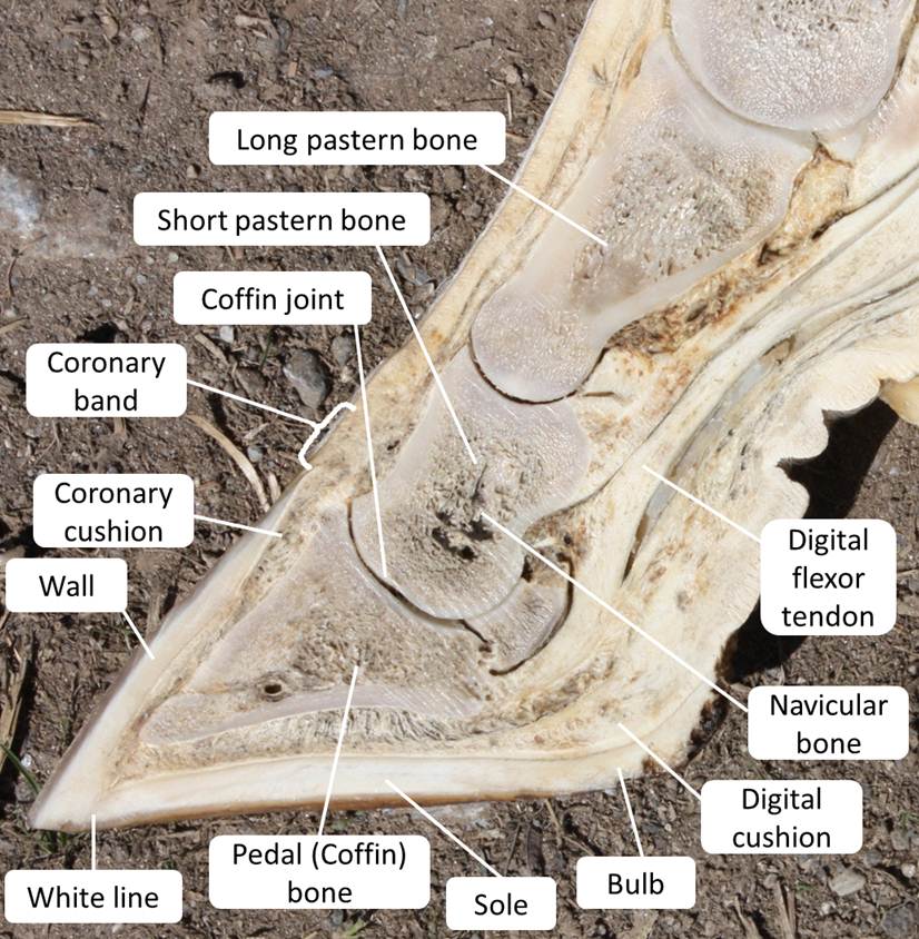
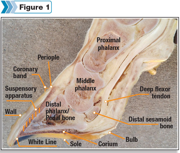
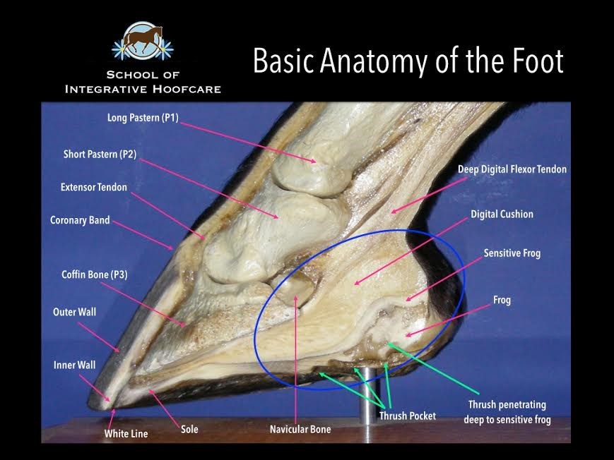
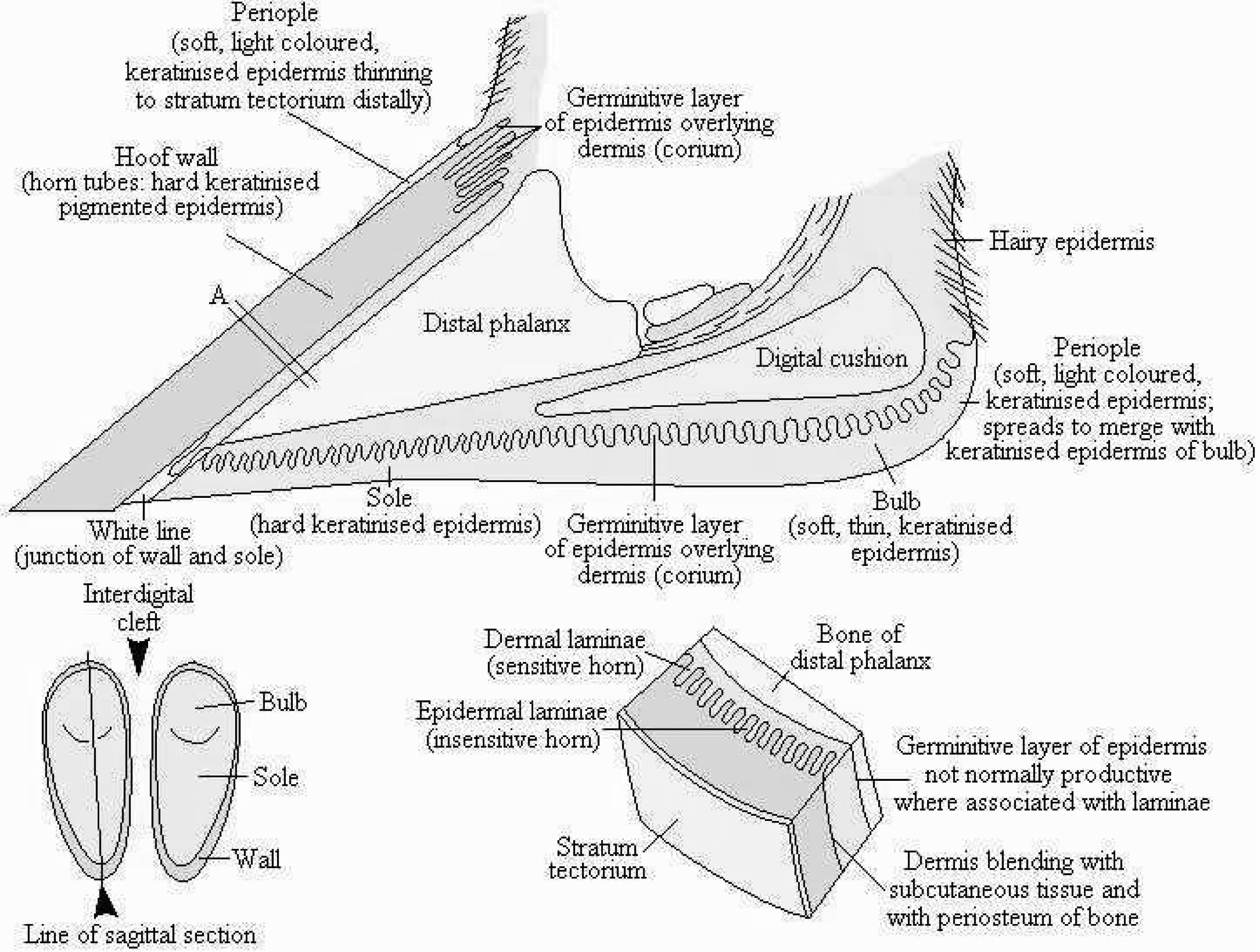
The hoof is defined from a physiologic perspective as the modified skin (epidermis) covering the tip of the digit and all enclosed structures. The hoof provides protection to the distal limb and is formed by keratinisation of the epithelial layer and modification of the underlying dermis.
Be proactive to prevent lameness during the dry period Progressive
Bovine Foot Anatomy Bones and joints While the cannon bone of a horse is MCIII or MTIII, in a cow it is a fused MCIII+IV or MTIII+IV. The fusion is present at the fetlock joint and above. Cattle do have the same bones and joints as horses below the fetlock but in duplicate form.

Cow Hoof Anatomy Model (2 part) 42010210 Anatomystuff
22/07/2023 28/07/2021 by Sonnet Cows are essential livestock that provides excellent value to their owner. The cow anatomy deals with the forms and structure of their particular organ. It is not possible to describe all the anatomical features of a cow in a single article.
cow hoof anatomy
Imaging Anatomy: Bovine Hindlimb Hoof Example 1 The following radiographs are the dorsoplantar, dorsolateral-plantaromedial oblique (DLPMO) and dorsomedial-plantarolateral oblique (DMPLO) views of the left rear foot of a nine-year-old Hereford cow. Click the radiograph image. Interactive image will open in a secondary window.
cow hoof anatomy
Knowing and understanding the anatomy of a cows hoof is absolutely essential in fully being able to take care of their hoof care - here I direct a hoof or di.
cow hoof anatomy
Vic's Hoof Trimming Course | Teach Anatomy Of Cattle Feet Legs. Vic's Hoof. About Us. +1 (519) 274-0878. (519) 679-6711.
PPT Chapter 4 PowerPoint Presentation, free download ID1114478
Lameness Wetlab: Hoof Anatomy and Basic Hoof Trimming Jason B. Osterstock, DVM, PhD Pfizer Animal Genetics, Kalamazoo, Ml 49007 Abstract Lameness is one of the most common causes of mor bidity in beef and dairy cattle operations and represents an important threat to the well-being of cattle in all types of operations.
Cow Foot Anatomy
The cow hoof anatomy is a cornified modification of the epidermis that lies under the vascular layer and covers the end of the digits. You will find two primary and two accessory hooves on each limb of a cow. The leading hoof of a cow comprises three parts - periople, wall, and sole.

The Dancing Donkey More alike than different
A healthy adult horse grows new hoof at the rate of between 1/4 to 1/2 inch per month. This means a horse will regrow its entire hoof wall over the course of about a year. Bovine hooves grow a bit slower, at a rate of approximately 2 inches a year. But, then again, a cow's hoof isn't as tall as a horse's hoof.

Bovine Hoof Anatomy
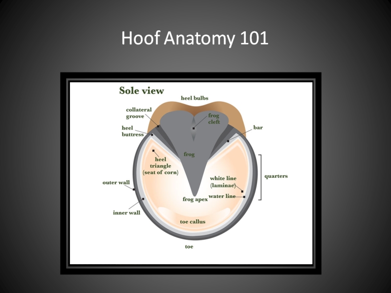
Cow Hoof Anatomy It's critical to distinguish between the various parts of the hoof, especially while foot trimming. Below is a detailed description of the nine parts of the hoof which are numbered in the picture above. Wall horn: Similar to human fingernails, this part is the strongest and most crucial for weight carrying.