
Diagram showing metatarsal bones of the foot. Download Scientific Diagram
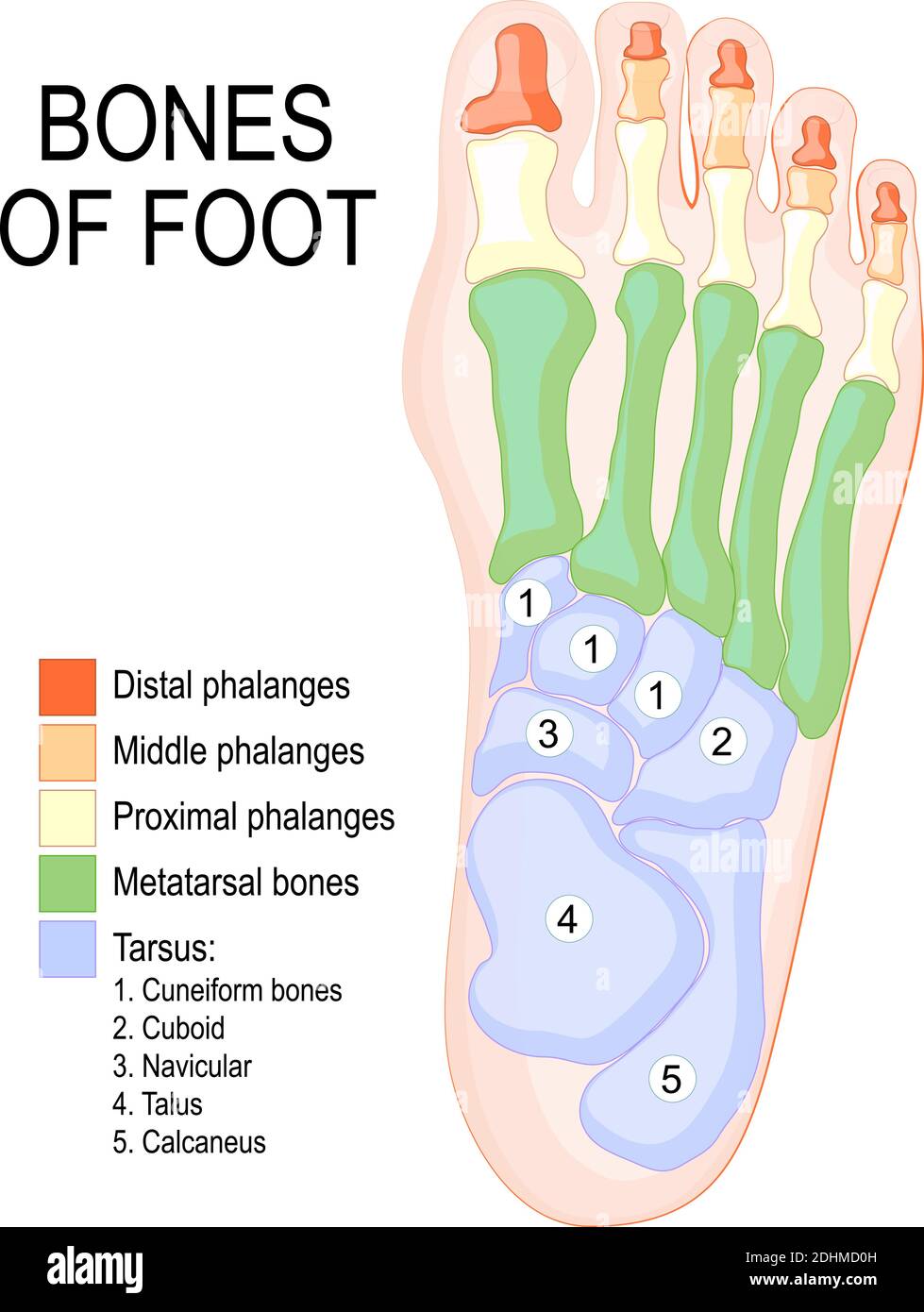
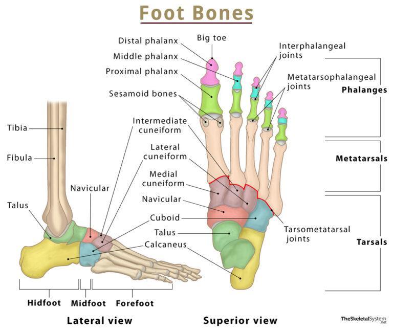
There are 26 bones in the foot, divided into three groups: Seven tarsal bones Five metatarsal bones Fourteen phalanges Tarsals make up a strong weight bearing platform. They are homologous to the carpals in the wrist and are divided into three groups: proximal, intermediate, and distal.
Bones of the human foot diagram 1142236 Vector Art at Vecteezy
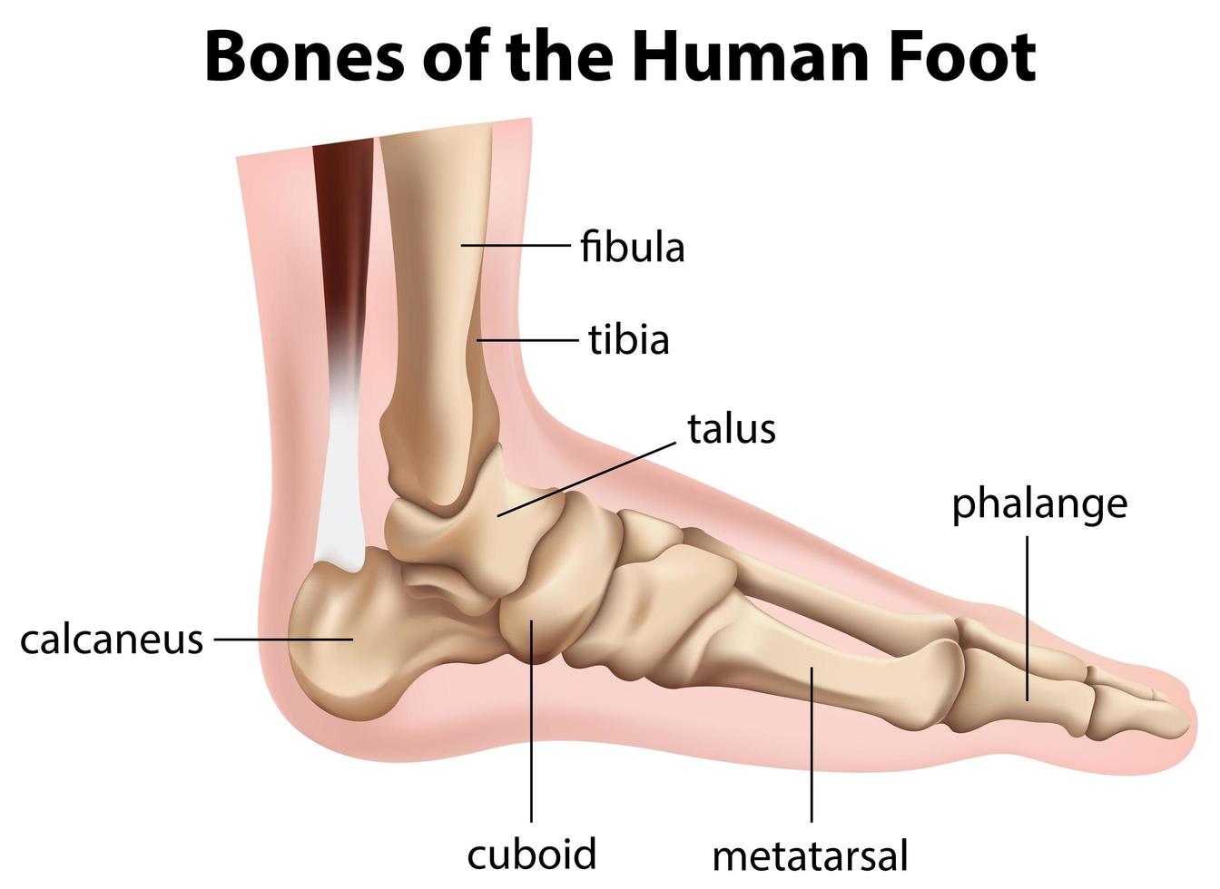
The foot can also be divided up into three regions: (i) Hindfoot - talus and calcaneus; (ii) Midfoot - navicular, cuboid, and cuneiforms; and (iii) Forefoot - metatarsals and phalanges. In this article, we shall look at the anatomy of the bones of the foot - their bony landmarks, articulations, and clinical correlations.
Pictures Of Bones Of The Feet
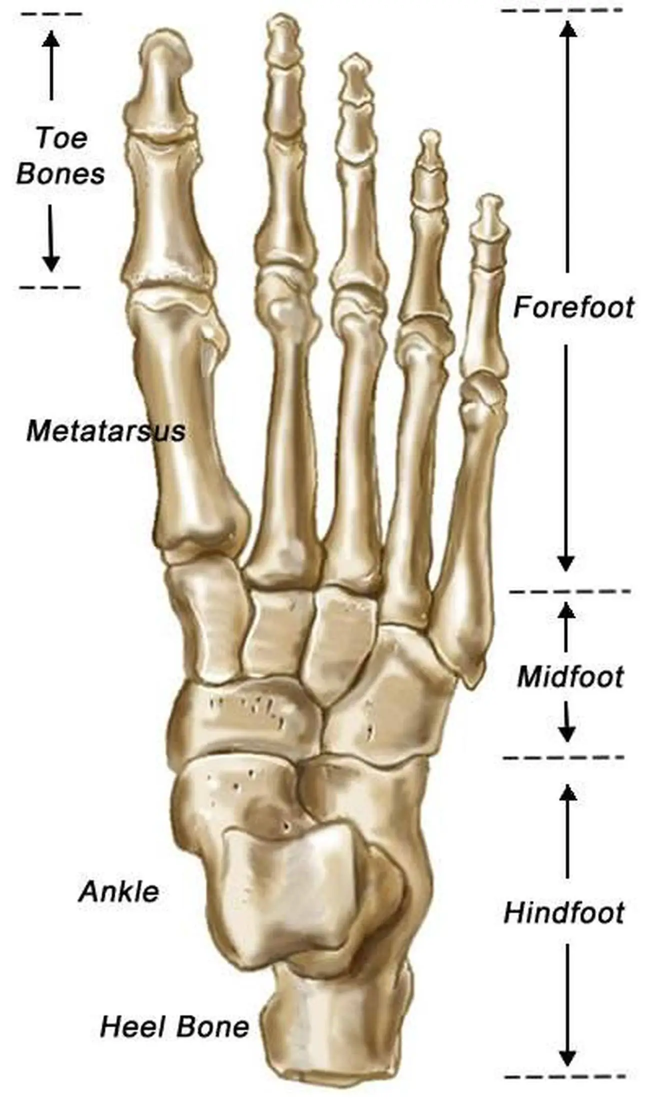
The foot itself can be divided into three sections: the hindfoot, midfoot and forefoot and the foot bones can be grouped into three sets: the tarsal bones, the metatarsals and the phalanges .

Ankle Bones Diagram . Ankle Bones Diagram Ankle Diagrams Diagram Link Ankle anatomy, Foot
The diagram of bones in the ankle and foot is given below: Tarsal Bones The tarsal bones in the foot are located amongst tibia, metatarsal bones, and fibula. There are in all 7 bones, which fall under tarsal bones category. They are: Calcaneus or Calcaneum: To explain the term in layman's language, it is the heel bone in the skeletal system.
Bones of foot. Human Anatomy. The diagram shows the placement and names of all bones of foot
The metatarsal bones are a group of five long bones located in the metatarsus of the foot, between the tarsal bones (near the ankle) and the phalanges (toe bones). These bones are numbered from one to five, starting with the first metatarsal beneath the big toe and moving laterally towards the fifth metatarsal beneath the little toe.
Anatomy The Bones Of The Foot
Details. The original file was in Wavefront .OBJ format. The following is the original legend from the file: Foot Bones # # Courtesy of: # # Viewpoint Animation Engineering # 870 West Center # Orem, Utah 84057 # (801)224-2222 # 1-800-DATASET # $ Contributed to the FTP site at avalon.chinalake.navy.mil (129.131.31.11) # by Scott R. Nelson of Sun.

Foot bones anatomy Royalty Free Vector Image VectorStock
The foot is the region of the body distal to the leg that is involved in weight bearing and locomotion. It consists of 28 bones, which can be divided functionally into three groups, referred to as the tarsus, metatarsus and phalanges. The foot is not only complicated in terms of the number and structure of bones, but also in terms of its joints.
.jpg)
Foot Bone Diagram resource Imageshare
Structure of the foot kool99/Getty Images In the foot, there are: 26 bones 33 joints more than 100 muscles, tendons, and ligaments Bones of the foot The bones in the foot make up.

Anatomy of the Foot and Ankle OrthoPaedia
Tibia Fibula Talus Cuneiforms Cuboid Navicular Many of the muscles that affect larger foot movements are located in the lower leg. However, the foot itself is a web of muscles that can perform.
Foot Bones Names, Anatomy, Structure, & Labeled Diagrams
Use these bones of the foot quizzes to master your identification skills. Overview of the bones of the foot and their divisions into the hindfoot, midfoot and forefoot. With a total of 26 bones in each foot, learning the bony anatomy of the foot is no piece of cake. That is, the memorization aspect.

Foot Description, Drawings, Bones, & Facts Britannica
Bones Of Foot Anatomy, Function & Diagram | Body Maps Human body Skeletal System Bones of foot Bones of foot The 26 bones of the foot consist of eight distinct types, including.

Foot Description, Drawings, Bones, & Facts Britannica
Fore-foot - the fore-foot is composed of the metatarsals and phalanges. The bones that comprise the fore-foot are those that are last to leave the ground during walking. Mobile Joints of the foot and ankle: (See Figure 3.) Ankle joint. Sub-talar joint. Talo-navicular joint. Metatarso-phalangeal (MTP) joints.

Bone Of Left Foot Anatomy Amp Physiology Illustration Human Anatomy Body
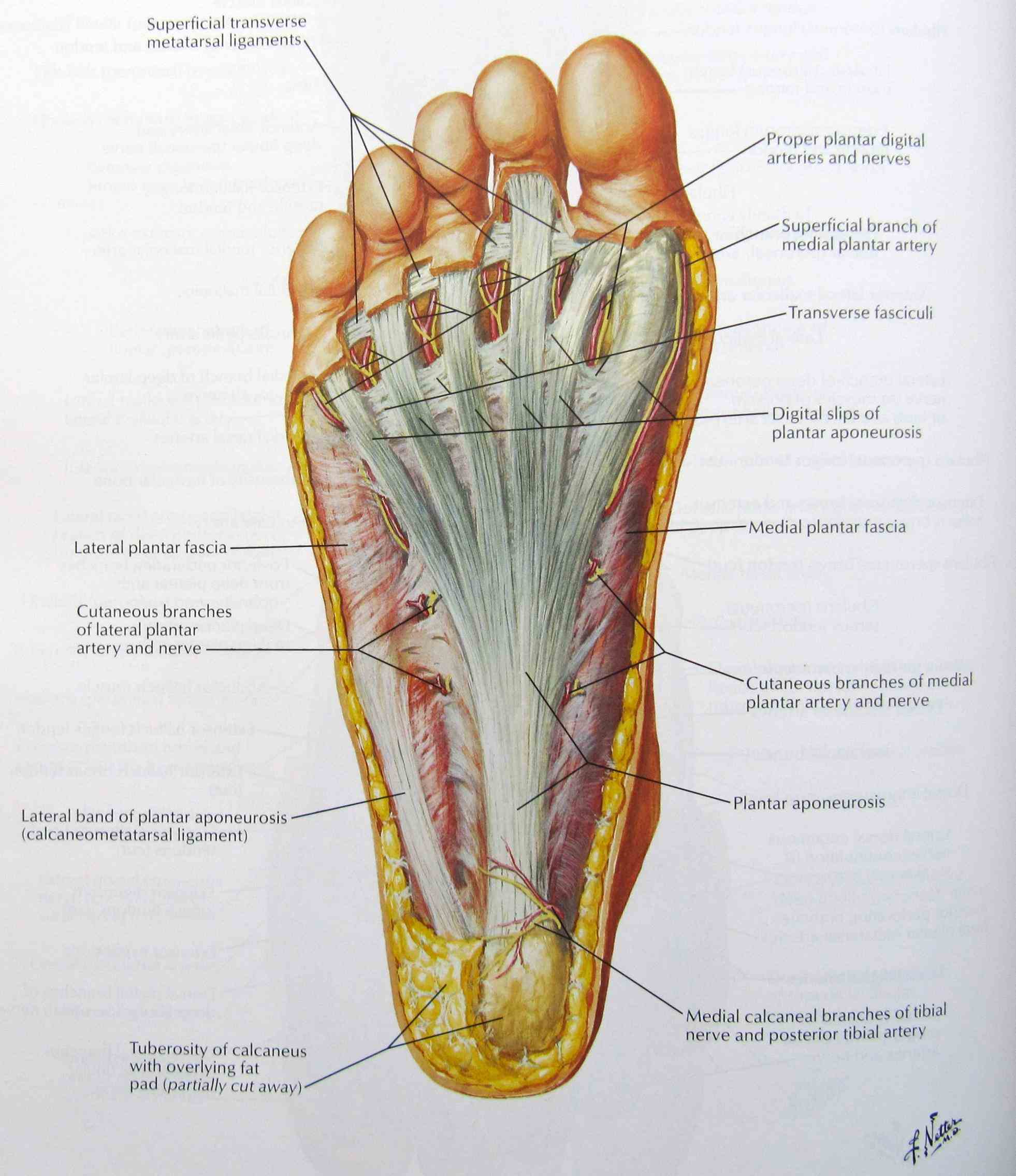
Introduction A solid understanding of anatomy is essential to effectively diagnose and treat patients with foot and ankle problems. Anatomy is a road map. Most structures in the foot are fairly superficial and can be easily palpated. Anatomical structures (tendons, bones, joints, etc) tend to hurt exactly where they are injured or inflamed.

Ankle and Foot Pain Massage Therapy Connections
Cuboid: This multi-faceted bone sits on the outside of the foot near the fifth phalanx (little toe). Cuneiforms: These three small bones are closest to the five metatarsal bones. They sit in a row.

Foot Bone Anatomy Vector Illustration 539973 Vector Art at Vecteezy
The anatomy of the foot The foot contains a lot of moving parts - 26 bones, 33 joints and over 100 ligaments. The foot is divided into three sections - the forefoot, the midfoot and the hindfoot. The forefoot This consists of five long bones (metatarsal bones) and five shorter bones that form the base of the toes (phalanges).
.jpg)
Foot Bone Diagram resource Imageshare
Summary The foot is an intricate part of the body, consisting of 26 bones, 33 joints, 107 ligaments, and 19 muscles. Scientists group the bones of the foot into the phalanges, tarsal.